2O2U

Crystal structure of human JNK3 complexed with N-(3-cyano-4,5,6,7-tetrahydro-1-benzothien-2-yl)-2-fluorobenzamide


Experimental Data Snapshot

  • Method: X-RAY DIFFRACTION
  • Resolution: 2.45 Å
  • R-Value Free: 0.281 
  • R-Value Work: 0.232 
  • R-Value Observed: 0.234 

wwPDB Validation   3D Report Full Report


Ligand Structure Quality Assessment 


This is version 1.4 of the entry. See complete history


Literature

N-(3-Cyano-4,5,6,7-tetrahydro-1-benzothien-2-yl)amides as potent, selective, inhibitors of JNK2 and JNK3.

Angell, R.M.Atkinson, F.L.Brown, M.J.Chuang, T.T.Christopher, J.A.Cichy-Knight, M.Dunn, A.K.Hightower, K.E.Malkakorpi, S.Musgrave, J.R.Neu, M.Rowland, P.Shea, R.L.Smith, J.L.Somers, D.O.Thomas, S.A.Thompson, G.Wang, R.

(2007) Bioorg Med Chem Lett 17: 1296-1301

  • DOI: https://doi.org/10.1016/j.bmcl.2006.12.003
  • Primary Citation of Related Structures:  
    2O0U, 2O2U

  • PubMed Abstract: 

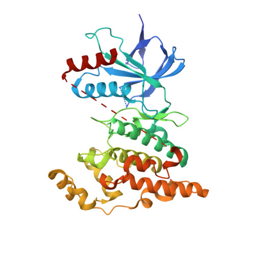
    The identification and exploration of a novel, potent and selective series of N-(3-cyano-4,5,6,7-tetrahydro-1-benzothien-2-yl)amide inhibitors of JNK2 and JNK3 kinases is described. Compounds 5a and 11a were identified as potent inhibitors of JNK3 (pIC50 6.7 and 6.6, respectively), with essentially equal potency against JNK2 (pIC50 6.5). Selectivity within the mitogen-activated protein kinase (MAPK) family, against JNK1, p38alpha and ERK2, was observed for the series. X-ray crystallography of 5e and 8a in JNK3 revealed a unique binding mode, with the 3-cyano substituent forming an H-bond acceptor interaction with the hinge region of the ATP-binding site.


  • Organizational Affiliation

    GlaxoSmithKline R&D, Medicines Research Centre, Gunnels Wood Road, Stevenage, Hertfordshire SG1 2NY, UK.


Macromolecules
Find similar proteins by:  (by identity cutoff)  |  3D Structure
Entity ID: 1
MoleculeChains Sequence LengthOrganismDetailsImage
Mitogen-activated protein kinase 10364Homo sapiensMutation(s): 0 
Gene Names: MAPK10JNK3JNK3APRKM10
EC: 2.7.11.24
UniProt & NIH Common Fund Data Resources
Find proteins for P53779 (Homo sapiens)
Explore P53779 
Go to UniProtKB:  P53779
PHAROS:  P53779
GTEx:  ENSG00000109339 
Entity Groups  
Sequence Clusters30% Identity50% Identity70% Identity90% Identity95% Identity100% Identity
UniProt GroupP53779
Sequence Annotations
Expand
  • Reference Sequence
Small Molecules
Ligands 1 Unique
IDChains Name / Formula / InChI Key2D Diagram3D Interactions
738
Query on 738

Download Ideal Coordinates CCD File 
B [auth A]N-(3-cyano-4,5,6,7-tetrahydro-1-benzothien-2-yl)-2-fluorobenzamide
C16 H13 F N2 O S
YVYPYORTKAIUGJ-UHFFFAOYSA-N
Binding Affinity Annotations 
IDSourceBinding Affinity
738 PDBBind:  2O2U Ki: 400 (nM) from 1 assay(s)
BindingDB:  2O2U IC50: min: 398.11, max: 1.00e+4 (nM) from 2 assay(s)
Binding MOAD:  2O2U IC50: 400 (nM) from 1 assay(s)
Experimental Data & Validation

Experimental Data

  • Method: X-RAY DIFFRACTION
  • Resolution: 2.45 Å
  • R-Value Free: 0.281 
  • R-Value Work: 0.232 
  • R-Value Observed: 0.234 
  • Space Group: P 21 21 21
Unit Cell:
Length ( Å )Angle ( ˚ )
a = 52.321α = 90
b = 71.376β = 90
c = 107.57γ = 90
Software Package:
Software NamePurpose
DENZOdata reduction
SCALEPACKdata scaling
REFMACrefinement
PDB_EXTRACTdata extraction
ADSCdata collection

Structure Validation

View Full Validation Report


Ligand Structure Quality Assessment 


Entry History 

Deposition Data

Revision History  (Full details and data files)

  • Version 1.0: 2007-02-27
    Type: Initial release
  • Version 1.1: 2008-05-01
    Changes: Version format compliance
  • Version 1.2: 2011-07-13
    Changes: Advisory, Version format compliance
  • Version 1.3: 2017-10-18
    Changes: Refinement description
  • Version 1.4: 2023-12-27
    Changes: Data collection, Database references, Derived calculations