2NZA

Structure and Function Studies of Cytochrome P450 158A1 from Streptomyces coelicolor A3(2)


Experimental Data Snapshot

  • Method: X-RAY DIFFRACTION
  • Resolution: 2.90 Å
  • R-Value Free: 0.267 
  • R-Value Work: 0.197 
  • R-Value Observed: 0.224 

wwPDB Validation   3D Report Full Report


Ligand Structure Quality Assessment 


This is version 1.3 of the entry. See complete history


Literature

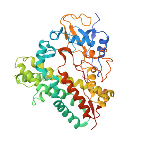
Different binding modes of two flaviolin substrate molecules in cytochrome P450 158A1 (CYP158A1) compared to CYP158A2.

Zhao, B.Lamb, D.C.Lei, L.Kelly, S.L.Yuan, H.Hachey, D.L.Waterman, M.R.

(2007) Biochemistry 46: 8725-8733

  • DOI: https://doi.org/10.1021/bi7006959
  • Primary Citation of Related Structures:  
    2DKK, 2NZ5, 2NZA

  • PubMed Abstract: 

    Cytochrome P450 158A2 (CYP158A2) has been shown to catalyze an unusual oxidative C-C coupling reaction to polymerize flaviolin and form highly conjugated pigments (three isomers of biflaviolin and one triflaviolin) in Streptomyces coelicolor A3(2) which protect the soil bacterium from deleterious effects of UV irradiation (Zhao B. et al. (2005) J. Biol. Chem. 280, 11599-11607). The present studies demonstrate that the subfamily partner CYP158A1, sharing 61% amino acid identity with CYP158A2, can also catalyze the same flaviolin dimerization reactions, but it generates just two of the three isomers of biflaviolin that CYP158A2 produces. Furthermore, the two CYP158A1 products have very different molar ratios compared with the corresponding CYP158A2 products, indicating that each enzyme maintains its own stereo- and regiospecificity. To find an explanation for these differences, three CYP158A1 structures have been solved by X-ray crystallography and have been compared with those for CYP158A2. The structures reveal surprising differences. Particularly, only one flaviolin molecule is present close to the heme iron in CYP158A1, and the second flaviolin molecule binds at the entrance of the putative substrate access channel on the protein distal surface 9 A away. Our work describes two members of the same P450 subfamily, which produce the same products by oxidative C-C coupling yet show very different structural orientations of substrate molecules in the active site.


  • Organizational Affiliation

    Department of Biochemistry, Vanderbilt University School of Medicine, Nashville, Tennessee 37232-0146, USA. bin.zhao@vanderbilt.edu


Macromolecules
Find similar proteins by:  (by identity cutoff)  |  3D Structure
Entity ID: 1
MoleculeChains Sequence LengthOrganismDetailsImage
Cytochrome P450 CYP158A1
A, B
413Streptomyces coelicolor A3(2)Mutation(s): 0 
Gene Names: SC8F11.24c
UniProt
Find proteins for Q9KZF5 (Streptomyces coelicolor (strain ATCC BAA-471 / A3(2) / M145))
Explore Q9KZF5 
Go to UniProtKB:  Q9KZF5
Entity Groups  
Sequence Clusters30% Identity50% Identity70% Identity90% Identity95% Identity100% Identity
UniProt GroupQ9KZF5
Sequence Annotations
Expand
  • Reference Sequence
Small Molecules
Ligands 1 Unique
IDChains Name / Formula / InChI Key2D Diagram3D Interactions
HEM
Query on HEM

Download Ideal Coordinates CCD File 
C [auth A],
D [auth B]
PROTOPORPHYRIN IX CONTAINING FE
C34 H32 Fe N4 O4
KABFMIBPWCXCRK-RGGAHWMASA-L
Experimental Data & Validation

Experimental Data

  • Method: X-RAY DIFFRACTION
  • Resolution: 2.90 Å
  • R-Value Free: 0.267 
  • R-Value Work: 0.197 
  • R-Value Observed: 0.224 
  • Space Group: C 1 2 1
Unit Cell:
Length ( Å )Angle ( ˚ )
a = 186.61α = 90
b = 44.348β = 98.23
c = 130.619γ = 90
Software Package:
Software NamePurpose
HKL-2000data collection
PHASERphasing
CNSrefinement
HKL-2000data reduction
HKL-2000data scaling

Structure Validation

View Full Validation Report


Ligand Structure Quality Assessment 


Entry History 

Deposition Data

Revision History  (Full details and data files)

  • Version 1.0: 2007-07-03
    Type: Initial release
  • Version 1.1: 2008-05-01
    Changes: Version format compliance
  • Version 1.2: 2011-07-13
    Changes: Source and taxonomy, Version format compliance
  • Version 1.3: 2023-08-30
    Changes: Data collection, Database references, Derived calculations, Refinement description