2NX9

Crystal structure of the carboxyltransferase domain of the oxaloacetate decarboxylase Na+ pump from Vibrio cholerae


Experimental Data Snapshot

  • Method: X-RAY DIFFRACTION
  • Resolution: 1.70 Å
  • R-Value Free: 0.284 
  • R-Value Work: 0.232 
  • R-Value Observed: 0.235 

wwPDB Validation   3D Report Full Report


This is version 1.3 of the entry. See complete history


Literature

Crystal Structure of the Carboxyltransferase Domain of the Oxaloacetate Decarboxylase Na(+) Pump from Vibrio cholerae.

Studer, R.Dahinden, P.Wang, W.W.Auchli, Y.Li, X.D.Dimroth, P.

(2007) J Mol Biol 367: 547-557

  • DOI: https://doi.org/10.1016/j.jmb.2006.12.035
  • Primary Citation of Related Structures:  
    2NX9

  • PubMed Abstract: 

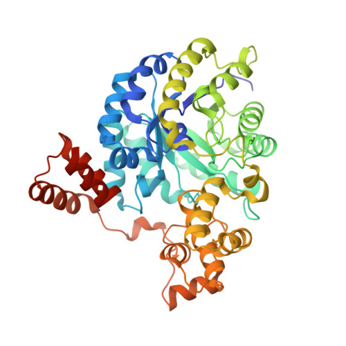
    Oxaloacetate decarboxylase is a membrane-bound multiprotein complex that couples oxaloacetate decarboxylation to sodium ion transport across the membrane. The initial reaction catalyzed by this enzyme machinery is the carboxyl transfer from oxaloacetate to the prosthetic biotin group. The crystal structure of the carboxyltransferase at 1.7 A resolution shows a dimer of alpha(8)beta(8) barrels with an active site metal ion, identified spectroscopically as Zn(2+), at the bottom of a deep cleft. The enzyme is completely inactivated by specific mutagenesis of Asp17, His207 and His209, which serve as ligands for the Zn(2+) metal ion, or by Lys178 near the active site, suggesting that Zn(2+) as well as Lys178 are essential for the catalysis. In the present structure this lysine residue is hydrogen-bonded to Cys148. A potential role of Lys178 as initial acceptor of the carboxyl group from oxaloacetate is discussed.


  • Organizational Affiliation

    Biomolecular Research, Paul Scherrer Institut, CH-5232 Villigen, Switzerland.


Macromolecules
Find similar proteins by:  (by identity cutoff)  |  3D Structure
Entity ID: 1
MoleculeChains Sequence LengthOrganismDetailsImage
Oxaloacetate decarboxylase 2, subunit alpha
A, B
464Vibrio choleraeMutation(s): 0 
Gene Names: oadA-2
EC: 4.1.1.3
UniProt
Find proteins for Q6A1F6 (Vibrio cholerae)
Explore Q6A1F6 
Go to UniProtKB:  Q6A1F6
Entity Groups  
Sequence Clusters30% Identity50% Identity70% Identity90% Identity95% Identity100% Identity
UniProt GroupQ6A1F6
Sequence Annotations
Expand
  • Reference Sequence
Experimental Data & Validation

Experimental Data

  • Method: X-RAY DIFFRACTION
  • Resolution: 1.70 Å
  • R-Value Free: 0.284 
  • R-Value Work: 0.232 
  • R-Value Observed: 0.235 
  • Space Group: P 21 21 21
Unit Cell:
Length ( Å )Angle ( ˚ )
a = 90.825α = 90
b = 91.655β = 90
c = 116.422γ = 90
Software Package:
Software NamePurpose
REFMACrefinement
XDSdata reduction
XSCALEdata scaling
MOLREPphasing

Structure Validation

View Full Validation Report


Entry History 

Deposition Data

Revision History  (Full details and data files)

  • Version 1.0: 2006-12-26
    Type: Initial release
  • Version 1.1: 2008-05-01
    Changes: Version format compliance
  • Version 1.2: 2011-07-13
    Changes: Advisory, Version format compliance
  • Version 1.3: 2023-08-30
    Changes: Data collection, Database references, Derived calculations, Refinement description