2NTU

Bacteriorhodopsin, wild type, before illumination


Experimental Data Snapshot

  • Method: X-RAY DIFFRACTION
  • Resolution: 1.53 Å
  • R-Value Free: 0.188 
  • R-Value Observed: 0.151 

wwPDB Validation   3D Report Full Report


Ligand Structure Quality Assessment 


This is version 1.3 of the entry. See complete history


Literature

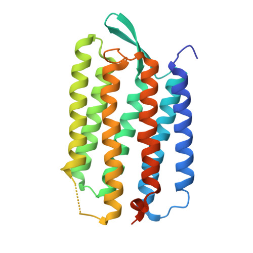
Structural changes in the L photointermediate of bacteriorhodopsin.

Lanyi, J.K.Schobert, B.

(2007) J Mol Biol 365: 1379-1392

  • DOI: https://doi.org/10.1016/j.jmb.2006.11.016
  • Primary Citation of Related Structures:  
    2NTU, 2NTW

  • PubMed Abstract: 

    The L to M reaction of the bacteriorhodopsin photocycle includes the crucial proton transfer from the retinal Schiff base to Asp85. In spite of the importance of the L state in deciding central issues of the transport mechanism in this pump, the serious disagreements among the three published crystallographic structures of L have remained unresolved. Here, we report on the X-ray diffraction structure of the L state, to 1.53-1.73 A resolutions, from replicate data sets collected from six independent crystals. Unlike earlier studies, the partial occupancy refinement uses diffraction intensities from the same crystals before and after the illumination to produce the trapped L state. The high reproducibility of inter-atomic distances, and bond angles and torsions of the retinal, lends credibility to the structural model. The photoisomerized 13-cis retinal in L is twisted at the C(13)=C(14) and C(15)=NZ double-bonds, and the Schiff base does not lose its connection to Wat402 and, therefore, to the proton acceptor Asp85. The protonation of Asp85 by the Schiff base in the L-->M reaction is likely to occur, therefore, via Wat402. It is evident from the structure of the L state that various conformational changes involving hydrogen-bonding residues and bound water molecules begin to propagate from the retinal to the protein at this stage already, and in both extracellular and cytoplasmic directions. Their rationales in the transport can be deduced from the way their amplitudes increase in the intermediates that follow L in the reaction cycle, and from the proton transfer reactions with which they are associated.


  • Organizational Affiliation

    Department of Physiology & Biophysics, University of California, Irvine, CA 92697, USA. jlanyi@orion.oac.uci.edu


Macromolecules
Find similar proteins by:  (by identity cutoff)  |  3D Structure
Entity ID: 1
MoleculeChains Sequence LengthOrganismDetailsImage
Bacteriorhodopsin249Halobacterium salinarumMutation(s): 0 
Gene Names: bop
Membrane Entity: Yes 
UniProt
Find proteins for P02945 (Halobacterium salinarum (strain ATCC 700922 / JCM 11081 / NRC-1))
Explore P02945 
Go to UniProtKB:  P02945
Entity Groups  
Sequence Clusters30% Identity50% Identity70% Identity90% Identity95% Identity100% Identity
UniProt GroupP02945
Sequence Annotations
Expand
  • Reference Sequence
Small Molecules
Ligands 1 Unique
IDChains Name / Formula / InChI Key2D Diagram3D Interactions
RET
Query on RET

Download Ideal Coordinates CCD File 
B [auth A]RETINAL
C20 H28 O
NCYCYZXNIZJOKI-OVSJKPMPSA-N
Experimental Data & Validation

Experimental Data

  • Method: X-RAY DIFFRACTION
  • Resolution: 1.53 Å
  • R-Value Free: 0.188 
  • R-Value Observed: 0.151 
  • Space Group: P 63
Unit Cell:
Length ( Å )Angle ( ˚ )
a = 60.966α = 90
b = 60.966β = 90
c = 110.388γ = 120
Software Package:
Software NamePurpose
SHELXmodel building
SHELXL-97refinement
ADSCdata collection
HKL-2000data reduction
HKL-2000data scaling
SHELXphasing

Structure Validation

View Full Validation Report


Ligand Structure Quality Assessment 


Entry History 

Deposition Data

Revision History  (Full details and data files)

  • Version 1.0: 2006-12-26
    Type: Initial release
  • Version 1.1: 2008-05-01
    Changes: Version format compliance
  • Version 1.2: 2011-07-13
    Changes: Derived calculations, Version format compliance
  • Version 1.3: 2023-08-30
    Changes: Data collection, Database references, Derived calculations, Refinement description