2NO4

Crystal Structure analysis of a Dehalogenase


Experimental Data Snapshot

  • Method: X-RAY DIFFRACTION
  • Resolution: 1.93 Å
  • R-Value Free: 0.216 
  • R-Value Work: 0.184 
  • R-Value Observed: 0.185 

wwPDB Validation   3D Report Full Report


This is version 1.4 of the entry. See complete history


Literature

Crystal structures of the substrate free-enzyme, and reaction intermediate of the HAD superfamily member, haloacid dehalogenase DehIVa from Burkholderia cepacia MBA4

Schmidberger, J.W.Wilce, J.A.Tsang, J.S.H.Wilce, M.C.J.

(2007) J Mol Biol 368: 706-717

  • DOI: https://doi.org/10.1016/j.jmb.2007.02.015
  • Primary Citation of Related Structures:  
    2NO4, 2NO5

  • PubMed Abstract: 

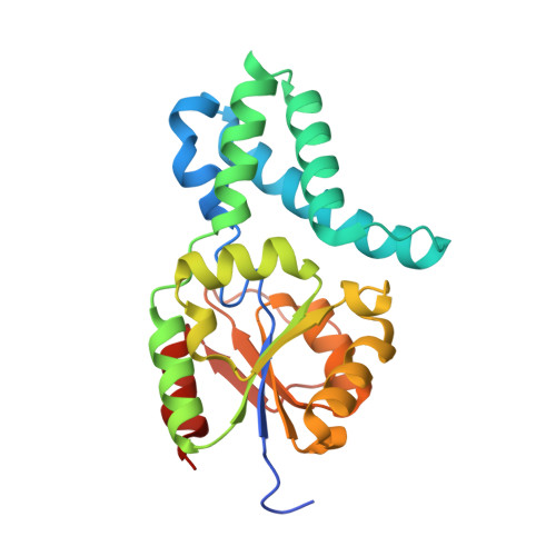
    DehIVa is a haloacid dehalogenase (EC 3.8.1.2) from the soil and water borne bacterium Burkholderia cepacia MBA4, which belongs to the functionally variable haloacid dehalogenase (HAD) superfamily of enzymes. The haloacid dehalogenases catalyse the removal of halides from haloacids resulting in a hydroxlated product. These enzymes are of interest for their potential to degrade recalcitrant halogenated environmental pollutants and their use in the synthesis of industrial chemicals. The haloacid dehalogenases utilise a nucleophilic attack on the substrate by an aspartic acid residue to form an enzyme-substrate ester bond and concomitantly cleaving of the carbon-halide bond and release of a hydroxylated product following ester hydrolysis. We present the crystal structures of both the substrate-free DehIVa refined to 1.93 A resolution and DehIVa covalently bound to l-2-monochloropropanoate trapped as a reaction intermediate, refined to 2.7 A resolution. Electron density consistent with a previously unidentified yet anticipated water molecule in the active site poised to donate its hydroxyl group to the product and its proton to the catalytic Asp11 is evident. It has been unclear how substrate enters the active site of this and related enzymes. The results of normal mode analysis (NMA) are presented and suggest a means whereby the predicted global dynamics of the enzyme allow for entry of the substrate into the active site. In the context of these results, the possible role of Arg42 and Asn178 in a "lock down" mechanism affecting active site access is discussed. In silico substrate docking of enantiomeric substrates has been examined in order to evaluate the enzymes enantioselectivity.


  • Organizational Affiliation

    School of Medicine and Pharmacology, The University of Western Australia, Perth, Australia.


Macromolecules
Find similar proteins by:  (by identity cutoff)  |  3D Structure
Entity ID: 1
MoleculeChains Sequence LengthOrganismDetailsImage
(S)-2-haloacid dehalogenase IVA
A, B
240Burkholderia cepaciaMutation(s): 0 
EC: 3.8.1.2
UniProt
Find proteins for Q51645 (Burkholderia cepacia)
Explore Q51645 
Go to UniProtKB:  Q51645
Entity Groups  
Sequence Clusters30% Identity50% Identity70% Identity90% Identity95% Identity100% Identity
UniProt GroupQ51645
Sequence Annotations
Expand
  • Reference Sequence
Small Molecules
Ligands 2 Unique
IDChains Name / Formula / InChI Key2D Diagram3D Interactions
SO4
Query on SO4

Download Ideal Coordinates CCD File 
C [auth A]
D [auth A]
E [auth A]
F [auth A]
H [auth B]
C [auth A],
D [auth A],
E [auth A],
F [auth A],
H [auth B],
I [auth B],
J [auth B],
K [auth B],
L [auth B],
M [auth B]
SULFATE ION
O4 S
QAOWNCQODCNURD-UHFFFAOYSA-L
CL
Query on CL

Download Ideal Coordinates CCD File 
G [auth A],
N [auth B]
CHLORIDE ION
Cl
VEXZGXHMUGYJMC-UHFFFAOYSA-M
Experimental Data & Validation

Experimental Data

  • Method: X-RAY DIFFRACTION
  • Resolution: 1.93 Å
  • R-Value Free: 0.216 
  • R-Value Work: 0.184 
  • R-Value Observed: 0.185 
  • Space Group: P 31 2 1
Unit Cell:
Length ( Å )Angle ( ˚ )
a = 103.999α = 90
b = 103.999β = 90
c = 134.811γ = 120
Software Package:
Software NamePurpose
DENZOdata reduction
SCALEPACKdata scaling
MOLREPphasing
REFMACrefinement
PDB_EXTRACTdata extraction
MAR345dtbdata collection

Structure Validation

View Full Validation Report


Entry History 

Deposition Data

Revision History  (Full details and data files)

  • Version 1.0: 2007-09-25
    Type: Initial release
  • Version 1.1: 2011-07-13
    Changes: Derived calculations, Version format compliance
  • Version 1.2: 2014-11-12
    Changes: Structure summary
  • Version 1.3: 2017-10-18
    Changes: Refinement description
  • Version 1.4: 2023-10-25
    Changes: Data collection, Database references, Refinement description