2NND

The Structural Identification of the Interaction Site and Functional State of RBP for its Membrane Receptor


Experimental Data Snapshot

  • Method: X-RAY DIFFRACTION
  • Resolution: 1.60 Å
  • R-Value Free: 0.227 
  • R-Value Work: 0.190 
  • R-Value Observed: 0.198 

wwPDB Validation   3D Report Full Report


Ligand Structure Quality Assessment 


This is version 1.3 of the entry. See complete history


Literature

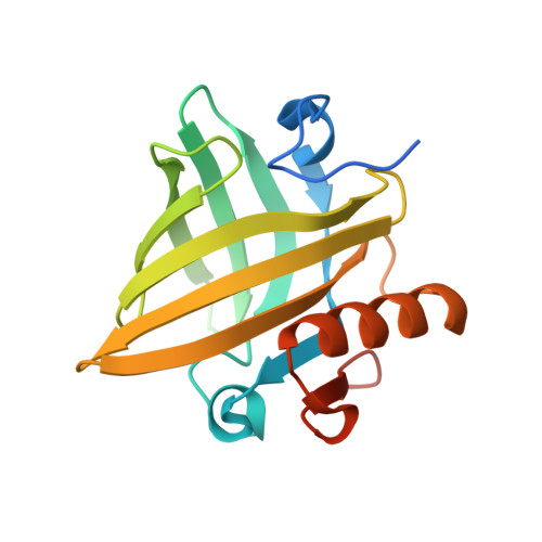
Identification of the retinol-binding protein (RBP) interaction site and functional state of RBPs for the membrane receptor.

Redondo, C.Vouropoulou, M.Evans, J.Findlay, J.B.

(2008) FASEB J 22: 1043-1054

  • DOI: https://doi.org/10.1096/fj.07-8939com
  • Primary Citation of Related Structures:  
    2NND, 2NNE

  • PubMed Abstract: 

    This laboratory has advanced a model whereby retinol is transported around the body bound to retinol-binding protein (RBP), is transferred across the membrane of cells by a specific receptor/transporter, and is picked up from the membrane by an intracellular homolog, cellular retinol-binding protein (CRBP). This process involves a number of protein-protein interactions, and we hypothesized that conformational changes were an integral part of the retinol transfer mechanism. Previously we identified the potential interaction site on RBP for its membrane receptor. Here we confirm by the analysis of chimera containing a grafted CD loop from RBP that this is indeed the receptor interaction site and go on to demonstrate that the conformational changes that occur to this region on the apo to holo transition in RBP also take place in a chimera binding a quite different ligand, thus establishing the concept. We have also gone on to support the hypothesis that CRBP may also bind to a receptor in the membrane. Previous evidence has indicated that one such receptor might be lecithin:retinol acyltransferase, an enzyme that catalyzes retinol esterification. Here we provide the first evidence that the plasma membrane receptor for RBP could be the same as that for CRBP. This observation offers support for the intracellular phase of the uptake process for retinol, providing an efficient and highly unique mechanism in eukaryotic biology.


  • Organizational Affiliation

    Institute of Membrane and Systems Biology, Faculty of Biological Sciences, LIGHT Laboratories, Clarendon Way, University of Leeds, Leeds LS2 9JT, UK.


Macromolecules
Find similar proteins by:  (by identity cutoff)  |  3D Structure
Entity ID: 1
MoleculeChains Sequence LengthOrganismDetailsImage
Major urinary protein 2178Mus musculusMutation(s): 0 
Gene Names: MUP1Mup2
UniProt
Find proteins for P02762 (Mus musculus)
Explore P02762 
Go to UniProtKB:  P02762
Entity Groups  
Sequence Clusters30% Identity50% Identity70% Identity90% Identity95% Identity100% Identity
UniProt GroupP02762
Sequence Annotations
Expand
  • Reference Sequence
Small Molecules
Binding Affinity Annotations 
IDSourceBinding Affinity
PRZ Binding MOAD:  2NND Kd: 870 (nM) from 1 assay(s)
PDBBind:  2NND Kd: 870 (nM) from 1 assay(s)
BindingDB:  2NND -TΔS: min: 9.39, max: 19.29 (kJ/mol) from 2 assay(s)
ΔG: min: -3.85e+1, max: -2.83e+1 (kJ/mol) from 2 assay(s)
Experimental Data & Validation

Experimental Data

  • Method: X-RAY DIFFRACTION
  • Resolution: 1.60 Å
  • R-Value Free: 0.227 
  • R-Value Work: 0.190 
  • R-Value Observed: 0.198 
  • Space Group: P 43 21 2
Unit Cell:
Length ( Å )Angle ( ˚ )
a = 53.636α = 90
b = 53.636β = 90
c = 137.347γ = 90
Software Package:
Software NamePurpose
REFMACrefinement
ADSCdata collection
MOSFLMdata reduction
SCALAdata scaling
CNSphasing

Structure Validation

View Full Validation Report


Ligand Structure Quality Assessment 


Entry History 

Deposition Data

Revision History  (Full details and data files)

  • Version 1.0: 2007-10-30
    Type: Initial release
  • Version 1.1: 2011-07-13
    Changes: Version format compliance
  • Version 1.2: 2017-08-23
    Changes: Source and taxonomy
  • Version 1.3: 2023-10-25
    Changes: Data collection, Database references, Derived calculations, Refinement description