2NLR

STREPTOMYCES LIVIDANS ENDOGLUCANASE (EC: 3.2.1.4) COMPLEX WITH MODIFIED GLUCOSE TRIMER


Experimental Data Snapshot

  • Method: X-RAY DIFFRACTION
  • Resolution: 1.20 Å
  • R-Value Free: 0.142 
  • R-Value Observed: 0.111 

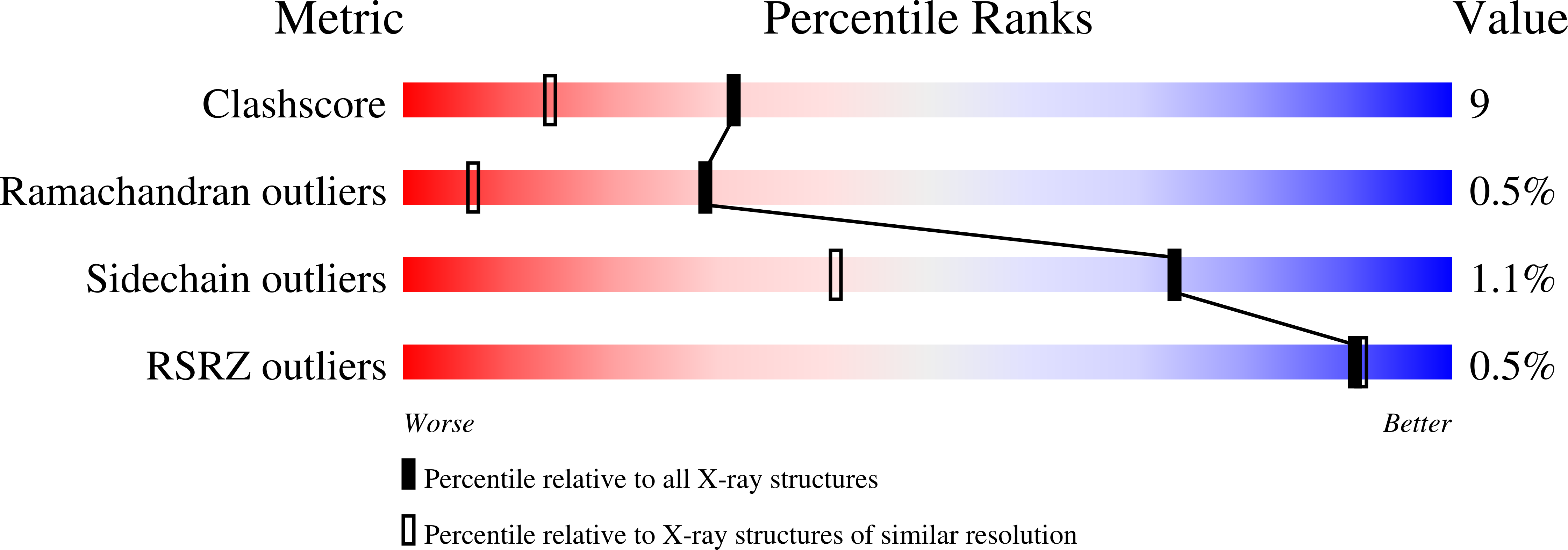
wwPDB Validation   3D Report Full Report


This is version 2.1 of the entry. See complete history


Literature

The crystal structure of a 2-fluorocellotriosyl complex of the Streptomyces lividans endoglucanase CelB2 at 1.2 A resolution.

Sulzenbacher, G.Mackenzie, L.F.Wilson, K.S.Withers, S.G.Dupont, C.Davies, G.J.

(1999) Biochemistry 38: 4826-4833

  • DOI: https://doi.org/10.1021/bi982648i
  • Primary Citation of Related Structures:  
    2NLR

  • PubMed Abstract: 

    Glycoside hydrolases have been classified into over 66 families on the basis of amino acid sequence. Recently a number of these families have been grouped into "clans" which share a common fold and catalytic mechanism [Henrissat, B., and Bairoch, A. (1996) Biochem. J. 316, 695-696]. Glycoside hydrolase Clan GH-C groups family 11 xylanases and family 12 cellulases, which share the same jellyroll topology, with two predominantly antiparallel beta-sheets forming a long substrate-binding cleft, and act with net retention of anomeric configuration. Here we present the three-dimensional structure of a family 12 endoglucanase, Streptomyces lividans CelB2, in complex with a 2-deoxy-2-fluorocellotrioside. Atomic resolution (1.2 A) data allow clear identification of two distinct species in the crystal. One is the glycosyl-enzyme intermediate, with the mechanism-based inhibitor covalently linked to the nucleophile Glu 120, and the other a complex with the reaction product, 2-deoxy-2-fluoro-beta-D-cellotriose. The active site architecture of the complex provides insight into the double-displacement mechanism of retaining glycoside hydrolases and also sheds light on the basis of the differences in specificity between family 12 cellulases and family 11 xylanases.


  • Organizational Affiliation

    Structural Biology Laboratory, Department of Chemistry, University of York, Heslington, York, Y010 5DD, U.K.


Macromolecules
Find similar proteins by:  (by identity cutoff)  |  3D Structure
Entity ID: 1
MoleculeChains Sequence LengthOrganismDetailsImage
PROTEIN (ENDOGLUCANASE (E.C.3.2.1.4))234Streptomyces lividansMutation(s): 0 
Gene Names: CELB
EC: 3.2.1.4
UniProt
Find proteins for Q54331 (Streptomyces lividans)
Explore Q54331 
Go to UniProtKB:  Q54331
Entity Groups  
Sequence Clusters30% Identity50% Identity70% Identity90% Identity95% Identity100% Identity
UniProt GroupQ54331
Sequence Annotations
Expand
  • Reference Sequence
Oligosaccharides

Help

Entity ID: 2
MoleculeChains Length2D Diagram Glycosylation3D Interactions
beta-D-glucopyranose-(1-4)-beta-D-glucopyranose-(1-4)-2-deoxy-2-fluoro-beta-D-glucopyranose
B
4N/A
Glycosylation Resources
GlyTouCan:  G98793XJ
GlyCosmos:  G98793XJ
Experimental Data & Validation

Experimental Data

  • Method: X-RAY DIFFRACTION
  • Resolution: 1.20 Å
  • R-Value Free: 0.142 
  • R-Value Observed: 0.111 
  • Space Group: P 31 2 1
Unit Cell:
Length ( Å )Angle ( ˚ )
a = 65.96α = 90
b = 65.96β = 90
c = 88.741γ = 120
Software Package:
Software NamePurpose
DENZOdata reduction
SCALEPACKdata scaling
AMoREphasing
SHELXLrefinement

Structure Validation

View Full Validation Report


Entry History 

Deposition Data

Revision History  (Full details and data files)

  • Version 1.0: 1999-11-10
    Type: Initial release
  • Version 1.1: 2007-10-16
    Changes: Version format compliance
  • Version 1.2: 2011-07-13
    Changes: Non-polymer description, Version format compliance
  • Version 1.3: 2017-10-04
    Changes: Refinement description
  • Version 2.0: 2020-07-29
    Type: Remediation
    Reason: Carbohydrate remediation
    Changes: Atomic model, Data collection, Derived calculations, Structure summary
  • Version 2.1: 2023-08-30
    Changes: Data collection, Database references, Refinement description, Structure summary