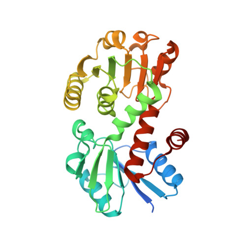
1.6 A structure of an NAD(+)-dependent quinate dehydrogenase from Corynebacterium glutamicum
Schoepe, J., Niefind, K., Schomburg, D.(2008) Acta Crystallogr D Biol Crystallogr 64: 803-809
- PubMed: 18566515
- DOI: https://doi.org/10.1107/S090744490801411X
- Primary Citation of Related Structures:
2NLO - PubMed Abstract:
To date, three different functional classes of bacterial shikimate/quinate dehydrogenases have been identified and are referred to as AroE, SDH-L and YdiB. The enzyme AroE and the catalytically much slower SDH-L clearly prefer NADP+/NADPH as the cosubstrate and are specific for (dehydro-)shikimate, whereas in YdiB the differences in affinity for NADP+/NADPH versus NAD+/NADH as well as for (dehydro-)shikimate versus (dehydro-)quinate are marginal. These three subclasses have a similar three-dimensional fold and hence all belong to the same structural class of proteins. In this paper, the crystal structure of an enzyme from Corynebacterium glutamicum is presented that clearly prefers NAD+ as a cosubstrate and that demonstrates a higher catalytic efficiency for quinate rather than shikimate. While the kinetic constants for this enzyme clearly differ from those reported for AroE, SDH-L and YdiB, the three-dimensional structure of this protein is similar to members of these three subclasses. Thus, the enzyme described here belongs to a new functional class of the shikimate/quinate dehydrogenase family. The different substrate and cosubstrate specificities of this enzyme relative to all other known bacterial shikimate/quinate dehydrogenases are discussed by means of analyzing the crystal structure and derived models. It is proposed that in contrast to shikimate, quinate forms a hydrogen bond to the NAD+. In addition, it is suggested that the hydroxyl group of a conserved active-site threonine hydrogen bonds to quinate more effectively than to shikimate. Also, the hydroxyl group of a conserved tyrosine approaches the carboxylate group of quinate more closely than it does the carboxylate group of shikimate. Taken together, these factors most likely lead to a lower Michaelis constant and therefore to a higher catalytic efficiency for quinate. The active site of the dehydrogenase reported here is larger than those of other known shikimate/quinate dehydrogenases, which may explain why quinate is easily accommodated within the catalytic cleft.
Organizational Affiliation:
Institute for Biochemistry, University of Cologne, Germany. j.schoepe@yahoo.de