Crystal structure of Myxococcus xanthus nucleoside diphosphate kinase and its interaction with a nucleotide substrate at 2.0 A resolution.
Williams, R.L., Oren, D.A., Munoz-Dorado, J., Inouye, S., Inouye, M., Arnold, E.(1993) J Mol Biol 234: 1230-1247
- PubMed: 8263923
- DOI: https://doi.org/10.1006/jmbi.1993.1673
- Primary Citation of Related Structures:
1NLK, 2NCK - PubMed Abstract:
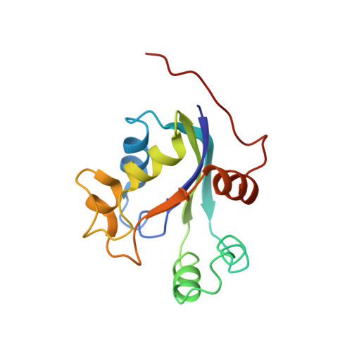
The X-ray crystallographic structure of nucleoside diphosphate (NDP) kinase from Myxococcus xanthus has been determined using multiple isomorphous replacement techniques and refined at 2.0 A resolution to a crystallographic R-factor of 0.17. This is the first report of the structure of an enzymatically active NDP kinase and of the enzyme with a bound nucleotide. The structure has been determined in P4(3)2(1)2 and I222 crystal forms. The enzyme monomer consists of a four-stranded antiparallel beta-sheet. The surfaces of the sheet are partially covered with five helical segments. There are two protein molecules in the asymmetric unit of the tetragonal crystal form. They form a dimer with an extensive interface in which 1092 A2 per monomer is buried. The majority of the contact area in the dimer interface is between hydrophobic or aromatic residues. Two dimers are related by a crystallographic 2-fold axis to yield a tetramer. This tetramer is also present in the orthorhombic crystals; however, in this case, the 222 symmetry is entirely crystallographic. Upon tetramer formation, an additional 473 A2 of solvent-accessible surface area from each monomer becomes buried. The interface between dimers in the tetramer is stabilized by salt bridges. Equilibrium sedimentation studies are consistent with the enzyme being a tetramer in solution. The structure of a complex of adenosine diphosphate (ADP) with the enzyme was determined and reveals that most of the nucleotide interactions with the protein are with the pyrophosphate and ribose groups, while the base has no hydrogen bonds with the protein and interacts only by stacking with the side chain of Phe59. The Mg2+ interacts with the pyrophosphate of the ADP and via a solvent molecule with the side chain of the conserved Asp120 residue. The mode of interaction with the nucleotide is novel, with the nucleotide binding at the side of the beta-sheet. The structures of the nucleotide in crystals grown in the presence or absence of Mg2+ are essentially identical. In addition, the phosphotransfer reaction from adenosine triphosphate (ATP) to the enzyme can occur without Mg2+. This suggests that only the second step of the reaction in which the enzyme transfers the phosphate to a nucleoside diphosphate acceptor is significantly catalyzed by the metal.
Organizational Affiliation:
Centre for Protein Engineering Medical Research Council Centre, Cambridge, England.