Dimeric switch of Hakai-truncated monomers during substrate recognition: insights from solution studies and NMR structure.
Mukherjee, M., Jing-Song, F., Ramachandran, S., Guy, G.R., Sivaraman, J.(2014) J Biol Chem 289: 25611-25623
- PubMed: 25074933
- DOI: https://doi.org/10.1074/jbc.M114.592840
- Primary Citation of Related Structures:
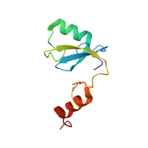
2MQ1 - PubMed Abstract:
Hakai, an E3 ubiquitin ligase, disrupts cell-cell contacts in epithelial cells and is up-regulated in human colon and gastric adenocarcinomas. Hakai acts through its phosphotyrosine-binding (HYB) domain, which bears a dimeric fold that recognizes the phosphotyrosine motifs of E-cadherin, cortactin, DOK1, and other Src substrates. Unlike the monomeric nature of the SH2 and phosphotyrosine-binding domains, the architecture of the HYB domain consists of an atypical, zinc-coordinated tight homodimer. Here, we report a C-terminal truncation mutant of the HYB domain (HYB(ΔC)), comprising amino acids 106-194, which exists as a monomer in solution. The NMR structure revealed that this deletion mutant undergoes a dramatic structural change caused by a rearrangement of the atypical zinc-coordinated unit in the C terminus of the HYB domain to a C2H2-like zinc finger in HYB(ΔC). Moreover, using isothermal titration calorimetry, we show that dimerization of HYB(ΔC) can be induced using a phosphotyrosine substrate peptide. This ligand-induced dimerization of HYB(ΔC) is further validated using analytical ultracentrifugation, size-exclusion chromatography, NMR relaxation studies, dynamic light scattering, and circular dichroism experiments. Overall, these observations suggest that the dimeric architecture of the HYB domain is essential for the phosphotyrosine-binding property of Hakai.
Organizational Affiliation:
From the Department of Biological Sciences, 14 Science Drive 4, National University of Singapore, Singapore 117543 and.