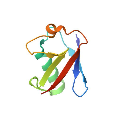
Unique structural, dynamical, and functional properties of k11-linked polyubiquitin chains.
Castaneda, C.A., Kashyap, T.R., Nakasone, M.A., Krueger, S., Fushman, D.(2013) Structure 21: 1168-1181
- PubMed: 23823328
- DOI: https://doi.org/10.1016/j.str.2013.04.029
- Primary Citation of Related Structures:
2MBO, 2MBQ - PubMed Abstract:
K11-linked polyubiquitin chains play important signaling and regulatory roles in both degradative and nonproteolytic pathways in eukaryotes. To understand the structural basis of how these chains are recognized and distinguished from other polyubiquitins, we determined solution structures of K11-linked diubiquitin (K11-Ub2) in the absence and presence of salt. These structures reveal that K11-Ub2 adopts conformations distinct from those of K48-linked or K63-linked chains. Importantly, our solution NMR and SANS data are inconsistent with published crystal structures of K11-Ub2. We found that increasing salt concentration compacts K11-Ub2 and strengthens interactions between the two Ub units. Binding studies indicate that K11-Ub2 interacts with ubiquitin-receptor proteins from both proteasomal and nonproteasomal pathways but with intermediate affinity and different binding modes than either K48-linked or K63-linked diubiquitin. Our data support the hypothesis that polyubiquitin chains of different linkages possess unique conformational and dynamical properties, allowing them to be recognized differently by downstream receptor proteins.
Organizational Affiliation:
Department of Chemistry and Biochemistry, Center for Biomolecular Structure and Organization, University of Maryland, College Park, MD 20742, USA.