2LGS

FEEDBACK INHIBITION OF FULLY UNADENYLYLATED GLUTAMINE SYNTHETASE FROM SALMONELLA TYPHIMURIUM BY GLYCINE, ALANINE, AND SERINE


Experimental Data Snapshot

  • Method: X-RAY DIFFRACTION
  • Resolution: 2.80 Å
  • R-Value Work: 0.235 
  • R-Value Observed: 0.235 

wwPDB Validation   3D Report Full Report


This is version 1.3 of the entry. See complete history


Literature

Feedback inhibition of fully unadenylylated glutamine synthetase from Salmonella typhimurium by glycine, alanine, and serine.

Liaw, S.H.Pan, C.Eisenberg, D.

(1993) Proc Natl Acad Sci U S A 90: 4996-5000

  • DOI: https://doi.org/10.1073/pnas.90.11.4996
  • Primary Citation of Related Structures:  
    2LGS

  • PubMed Abstract: 

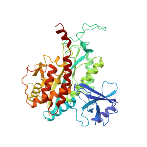
    Bacterial glutamine synthetase (GS; EC 6.3.1.2) was previously shown to be inhibited by nine end products of glutamine metabolism. Here we present four crystal structures of GS, complexed with the substrate Glu and with each of three feedback inhibitors. The GS of the present study is from Salmonella typhimurium, with Mn2+ ions bound, and is fully unadenylylated. From Fourier difference maps, we find that L-serine, L-alanine, and glycine bind at the site of the substrate L-glutamate. In our model, these four amino acids bind with the atoms they share in common (the "main chain" +NH3-CH-COO-) in the same positions. Thus on the basis of our x-ray work, glycine, alanine, and serine appear to inhibit GS-Mn by competing with the substrate glutamate for the active site.


  • Organizational Affiliation

    Molecular Biology Institute, University of California, Los Angeles 90024.


Macromolecules
Find similar proteins by:  (by identity cutoff)  |  3D Structure
Entity ID: 1
MoleculeChains Sequence LengthOrganismDetailsImage
GLUTAMINE SYNTHETASE
A, B, C, D, E
A, B, C, D, E, F, G, H, I, J, K, L
468Salmonella enterica subsp. enterica serovar TyphimuriumMutation(s): 0 
EC: 6.3.1.2
UniProt
Find proteins for P0A1P6 (Salmonella typhimurium (strain LT2 / SGSC1412 / ATCC 700720))
Explore P0A1P6 
Go to UniProtKB:  P0A1P6
Entity Groups  
Sequence Clusters30% Identity50% Identity70% Identity90% Identity95% Identity100% Identity
UniProt GroupP0A1P6
Sequence Annotations
Expand
  • Reference Sequence
Small Molecules
Ligands 2 Unique
IDChains Name / Formula / InChI Key2D Diagram3D Interactions
GLU
Query on GLU

Download Ideal Coordinates CCD File 
AA [auth E]
DA [auth F]
GA [auth G]
JA [auth H]
MA [auth I]
AA [auth E],
DA [auth F],
GA [auth G],
JA [auth H],
MA [auth I],
O [auth A],
PA [auth J],
R [auth B],
SA [auth K],
U [auth C],
VA [auth L],
X [auth D]
GLUTAMIC ACID
C5 H9 N O4
WHUUTDBJXJRKMK-VKHMYHEASA-N
MN
Query on MN

Download Ideal Coordinates CCD File 
BA [auth F]
CA [auth F]
EA [auth G]
FA [auth G]
HA [auth H]
BA [auth F],
CA [auth F],
EA [auth G],
FA [auth G],
HA [auth H],
IA [auth H],
KA [auth I],
LA [auth I],
M [auth A],
N [auth A],
NA [auth J],
OA [auth J],
P [auth B],
Q [auth B],
QA [auth K],
RA [auth K],
S [auth C],
T [auth C],
TA [auth L],
UA [auth L],
V [auth D],
W [auth D],
Y [auth E],
Z [auth E]
MANGANESE (II) ION
Mn
WAEMQWOKJMHJLA-UHFFFAOYSA-N
Experimental Data & Validation

Experimental Data

  • Method: X-RAY DIFFRACTION
  • Resolution: 2.80 Å
  • R-Value Work: 0.235 
  • R-Value Observed: 0.235 
  • Space Group: C 1 2 1
Unit Cell:
Length ( Å )Angle ( ˚ )
a = 235.5α = 90
b = 134.5β = 102.8
c = 200.1γ = 90
Software Package:
Software NamePurpose
X-PLORmodel building
X-PLORrefinement
X-PLORphasing

Structure Validation

View Full Validation Report


Entry History 

Deposition Data

Revision History  (Full details and data files)

  • Version 1.0: 1994-11-30
    Type: Initial release
  • Version 1.1: 2008-03-03
    Changes: Version format compliance
  • Version 1.2: 2011-07-13
    Changes: Version format compliance
  • Version 1.3: 2024-02-21
    Changes: Data collection, Database references, Derived calculations, Other