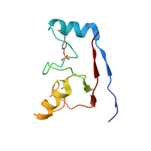
Autoinhibition and phosphorylation-induced activation mechanisms of human cancer and autoimmune disease-related E3 protein Cbl-b
Kobashigawa, Y., Tomitaka, A., Kumeta, H., Noda, N.N., Yamaguchi, M., Inagaki, F.(2011) Proc Natl Acad Sci U S A 108: 20579-20584
- PubMed: 22158902
- DOI: https://doi.org/10.1073/pnas.1110712108
- Primary Citation of Related Structures:
2LDR, 3VGO - PubMed Abstract:
Cbl-b is a RING-type E3 ubiquitin ligase that functions as a negative regulator of T-cell activation and growth factor receptor and nonreceptor-type tyrosine kinase signaling. Cbl-b dysfunction is related to autoimmune diseases and cancers in humans. However, the molecular mechanism regulating its E3 activity is largely unknown. NMR and small-angle X-ray scattering analyses revealed that the unphosphorylated N-terminal region of Cbl-b forms a compact structure by an intramolecular interaction, which masks the interaction surface of the RING domain with an E2 ubiquitin-conjugating enzyme. Phosphorylation of Y363, located in the helix-linker region between the tyrosine kinase binding and the RING domains, disrupts the interdomain interaction to expose the E2 binding surface of the RING domain. Structural analysis revealed that the phosphorylated helix-RING region forms a compact structure in solution. Moreover, the phosphate group of pY363 is located in the vicinity of the interaction surface with UbcH5B to increase affinity by reducing their electrostatic repulsion. Thus, the phosphorylation of Y363 regulates the E3 activity of Cbl-b by two mechanisms: one is to remove the masking of the RING domain from the tyrosine kinase binding domain and the other is to form a surface to enhance binding affinity to E2.
Organizational Affiliation:
Department of Structural Biology, Faculty of Advanced Life Science, and Graduate School of Life Science, Hokkaido University, Sapporo, Hokkaido 001-0021, Japan.