Probing the relationship between Gram-negative and Gram-positive S1 proteins by sequence analysis
Salah, P., Bisaglia, M., Aliprandi, P., Uzan, M., Sizun, C., Bontems, F.(2009) Nucleic Acids Res 37: 5578-5588
- PubMed: 19605565
- DOI: https://doi.org/10.1093/nar/gkp547
- Primary Citation of Related Structures:
2KHI, 2KHJ - PubMed Abstract:
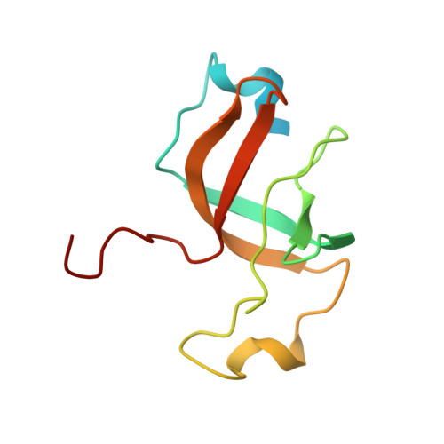
Escherichia coli ribosomal protein S1 is required for the translation initiation of messenger RNAs, in particular when their Shine-Dalgarno sequence is degenerated. Closely related forms of the protein, composed of the same number of domains (six), are found in all Gram-negative bacteria. More distant proteins, generally formed of fewer domains, have been identified, by sequence similarities, in Gram-positive bacteria and are also termed 'S1 proteins'. However in the absence of functional information, it is generally difficult to ascertain their relationship with Gram-negative S1. In this article, we report the solution structure of the fourth and sixth domains of the E. coli protein S1 and show that it is possible to characterize their beta-barrel by a consensus sequence that allows a precise identification of all domains in Gram-negative and Gram-positive S1 proteins. In addition, we show that it is possible to discriminate between five domain types corresponding to the domains 1, 2, 3, 4-5 and 6 of E. coli S1 on the basis of their sequence. This enabled us to identify the nature of the domains present in Gram-positive proteins and, subsequently, to probe the filiations between all forms of S1.
Organizational Affiliation:
CNRS, Centre de Recherche CNRS de Gif-sur-Yvette FRC 3115, Institut de Chimie des Substances Naturelles, 91198 Gif-sur-Yvette Cedex, France.