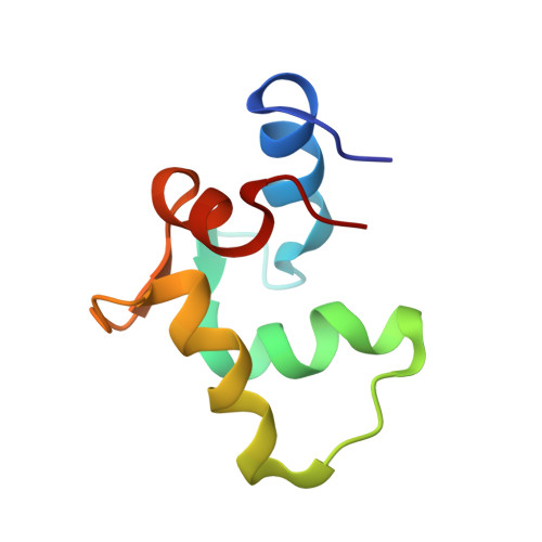
Solution structure of human cardiac troponin C in complex with the green tea polyphenol, (-)-epigallocatechin 3-gallate
Robertson, I.M., Li, M.X., Sykes, B.D.(2009) J Biol Chem 284: 23012-23023
- PubMed: 19542563
- DOI: https://doi.org/10.1074/jbc.M109.021352
- Primary Citation of Related Structures:
2KDH - PubMed Abstract:
Heart muscle contraction is regulated by Ca(2+) binding to the thin filament protein troponin C. In cardiovascular disease, the myofilament response to Ca(2+) is often altered. Compounds that rectify this perturbation are of considerable interest as therapeutics. Plant flavonoids have been found to provide protection against a variety of human illnesses such as cancer, infection, and heart disease. (-)-Epigallocatechin gallate (EGCg), the prevalent flavonoid in green tea, modulates force generation in isolated guinea pig hearts (Hotta, Y., Huang, L., Muto, T., Yajima, M., Miyazeki, K., Ishikawa, N., Fukuzawa, Y., Wakida, Y., Tushima, H., Ando, H., and Nonogaki, T. (2006) Eur. J. Pharmacol. 552, 123-130) and in skinned cardiac muscle fibers (Liou, Y. M., Kuo, S. C., and Hsieh, S. R. (2008) Pflugers Arch. 456, 787-800; and Tadano, N., Yumoto, F., Tanokura, M., Ohtsuki, I., and Morimoto, S. (2005) Biophys. J. 88, 314a). In this study we describe the solution structure of the Ca(2+)-saturated C-terminal domain of troponin C in complex with EGCg. Moreover, we show that EGCg forms a ternary complex with the C-terminal domain of troponin C and the anchoring region of troponin I. The structural evidence indicates that the binding site of EGCg on the C-terminal domain of troponin C is in the hydrophobic pocket in the absence of troponin I, akin to EMD 57033. Based on chemical shift mapping, the binding of EGCg to the C-terminal domain of troponin C in the presence of troponin I may be to a new site formed by the troponin C.troponin I complex. This interaction of EGCg with the C-terminal domain of troponin C.troponin I complex has not been shown with other cardiotonic molecules and illustrates the potential mechanism by which EGCg modulates heart contraction.
Organizational Affiliation:
Department of Biochemistry, University of Alberta, Edmonton, Alberta T6G 2H7, Canada.