2KAU

THE CRYSTAL STRUCTURE OF UREASE FROM KLEBSIELLA AEROGENES AT 2.2 ANGSTROMS RESOLUTION


Experimental Data Snapshot

  • Method: X-RAY DIFFRACTION
  • Resolution: 2.00 Å
  • R-Value Free: 0.225 
  • R-Value Work: 0.185 
  • R-Value Observed: 0.185 

wwPDB Validation   3D Report Full Report


This is version 1.2 of the entry. See complete history


Literature

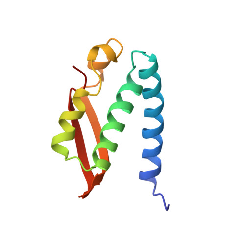
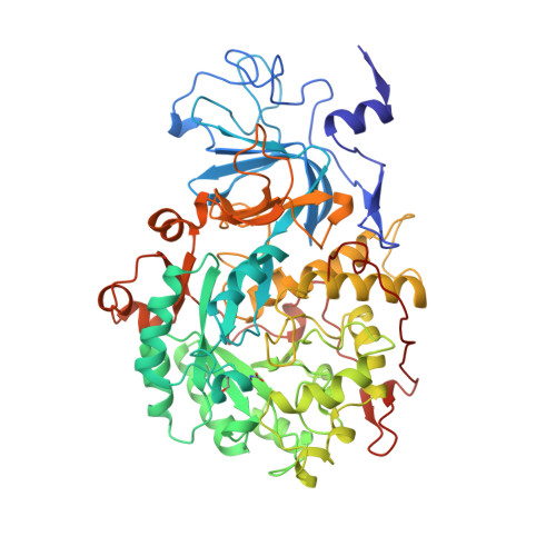
The crystal structure of urease from Klebsiella aerogenes.

Jabri, E.Carr, M.B.Hausinger, R.P.Karplus, P.A.

(1995) Science 268: 998-1004

  • Primary Citation of Related Structures:  
    2KAU

  • PubMed Abstract: 

    The crystal structure of urease from Klebsiella aerogenes has been determined at 2.2 A resolution and refined to an R factor of 18.2 percent. The enzyme contains four structural domains: three with novel folds playing structural roles, and an (alpha beta)8 barrel domain, which contains the bi-nickel center. The two active site nickels are 3.5 A apart. One nickel ion is coordinated by three ligands (with low occupancy of a fourth ligand) and the second is coordinated by five ligands. A carbamylated lysine provides an oxygen ligand to each nickel, explaining why carbon dioxide is required for the activation of urease apoenzyme. The structure is compatible with a catalytic mechanism whereby urea ligates Ni-1 to complete its tetrahedral coordination and a hydroxide ligand of Ni-2 attacks the carbonyl carbon. A surprisingly high structural similarity between the urease catalytic domain and that of the zinc-dependent adenosine deaminase reveals a remarkable example of active site divergence.


  • Organizational Affiliation

    Section of Biochemistry, Molecular and Cell Biology, Cornell University, Ithaca, NY 14853, USA.


Macromolecules
Find similar proteins by:  (by identity cutoff)  |  3D Structure
Entity ID: 1
MoleculeChains Sequence LengthOrganismDetailsImage
UREASE (GAMMA CHAIN)100Klebsiella aerogenesMutation(s): 0 
Gene Names: UREC
EC: 3.5.1.5
UniProt
Find proteins for P18316 (Klebsiella aerogenes)
Explore P18316 
Go to UniProtKB:  P18316
Entity Groups  
Sequence Clusters30% Identity50% Identity70% Identity90% Identity95% Identity100% Identity
UniProt GroupP18316
Sequence Annotations
Expand
  • Reference Sequence
Find similar proteins by:  (by identity cutoff)  |  3D Structure
Entity ID: 2
MoleculeChains Sequence LengthOrganismDetailsImage
UREASE (BETA CHAIN)106Klebsiella aerogenesMutation(s): 0 
Gene Names: UREB
EC: 3.5.1.5
UniProt
Find proteins for P18315 (Klebsiella aerogenes)
Explore P18315 
Go to UniProtKB:  P18315
Entity Groups  
Sequence Clusters30% Identity50% Identity70% Identity90% Identity95% Identity100% Identity
UniProt GroupP18315
Sequence Annotations
Expand
  • Reference Sequence
Find similar proteins by:  (by identity cutoff)  |  3D Structure
Entity ID: 3
MoleculeChains Sequence LengthOrganismDetailsImage
UREASE (ALPHA CHAIN)567Klebsiella aerogenesMutation(s): 0 
Gene Names: UREA
EC: 3.5.1.5
UniProt
Find proteins for P18314 (Klebsiella aerogenes)
Explore P18314 
Go to UniProtKB:  P18314
Entity Groups  
Sequence Clusters30% Identity50% Identity70% Identity90% Identity95% Identity100% Identity
UniProt GroupP18314
Sequence Annotations
Expand
  • Reference Sequence
Small Molecules
Modified Residues  1 Unique
IDChains TypeFormula2D DiagramParent
KCX
Query on KCX
C
L-PEPTIDE LINKINGC7 H14 N2 O4LYS
Experimental Data & Validation

Experimental Data

  • Method: X-RAY DIFFRACTION
  • Resolution: 2.00 Å
  • R-Value Free: 0.225 
  • R-Value Work: 0.185 
  • R-Value Observed: 0.185 
  • Space Group: I 21 3
Unit Cell:
Length ( Å )Angle ( ˚ )
a = 170.8α = 90
b = 170.8β = 90
c = 170.8γ = 90
Software Package:
Software NamePurpose
X-PLORmodel building
X-PLORrefinement
SDMSdata reduction
X-PLORphasing

Structure Validation

View Full Validation Report


Entry History 

Deposition Data

Revision History  (Full details and data files)

  • Version 1.0: 1995-07-10
    Type: Initial release
  • Version 1.1: 2008-03-03
    Changes: Version format compliance
  • Version 1.2: 2011-07-13
    Changes: Derived calculations, Version format compliance