Left-Handed Dimer of EphA2 Transmembrane Domain: Helix Packing Diversity among Receptor Tyrosine Kinases
Bocharov, E.V., Mayzel, M.L., Volynsky, P.E., Mineev, K.S., Tkach, E.N., Ermolyuk, Y.S., Schulga, A.A., Efremov, R.G., Arseniev, A.S.(2010) Biophys J 98: 881-889
- PubMed: 20197042
- DOI: https://doi.org/10.1016/j.bpj.2009.11.008
- Primary Citation of Related Structures:
2K9Y - PubMed Abstract:
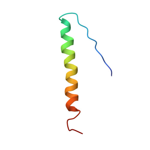
The Eph receptor tyrosine kinases and their membrane-bound ephrin ligands control a diverse array of cell-cell interactions in the developing and adult organisms. During signal transduction across plasma membrane, Eph receptors, like other receptor tyrosine kinases, are involved in lateral dimerization and subsequent oligomerization presumably with proper assembly of their single-span transmembrane domains. Spatial structure of dimeric transmembrane domain of EphA2 receptor embedded into lipid bicelle was obtained by solution NMR, showing a left-handed parallel packing of the transmembrane helices (535-559)(2). The helices interact through the extended heptad repeat motif L(535)X(3)G(539)X(2)A(542)X(3)V(546)X(2)L(549) assisted by intermolecular stacking interactions of aromatic rings of (FF(557))(2), whereas the characteristic tandem GG4-like motif A(536)X(3)G(540)X(3)G(544) is not used, enabling another mode of helix-helix association. Importantly, a similar motif AX(3)GX(3)G as was found is responsible for right-handed dimerization of transmembrane domain of the EphA1 receptor. These findings serve as an instructive example of the diversity of transmembrane domain formation within the same family of protein kinases and seem to favor the assumption that the so-called rotation-coupled activation mechanism may take place during the Eph receptor signaling. A possible role of membrane lipid rafts in relation to Eph transmembrane domain oligomerization and Eph signal transduction was also discussed.
Organizational Affiliation:
Shemyakin-Ovchinnikov Institute of Bioorganic Chemistry RAS, Moscow, Russia.