Solution structures of the trihelix DNA-binding domains of the wild-type and a phosphomimetic mutant of Arabidopsis GT-1: mechanism for an increase in DNA-binding affinity through phosphorylation.
Nagata, T., Niyada, E., Fujimoto, N., Nagasaki, Y., Noto, K., Miyanoiri, Y., Murata, J., Hiratsuka, K., Katahira, M.(2010) Proteins 78: 3033-3047
- PubMed: 20717979
- DOI: https://doi.org/10.1002/prot.22827
- Primary Citation of Related Structures:
2EBI, 2JMW - PubMed Abstract:
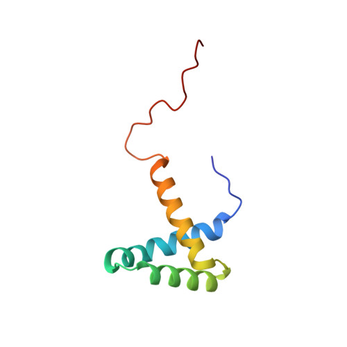
GT-1 is a plant transcription factor that binds to one of the cis-acting elements, BoxII, which resides within the upstream promoter region of light-responsive genes. GT-1 was assumed to act as a molecular switch modulated through Ca(2+)-dependent phosphorylation/dephosphorylation in response to light signals. It was shown previously that the phosphorylation of threonine 133 in the DNA-binding domain (DBD) of GT-1 results in enhancement of the BoxII-binding activity. Interestingly, point mutation of Thr133 to Asp also enhances the BoxII-binding activity. Here, we report the solution structures of hypothetical trihelix DBDs of the wild-type (WT) and a phosphomimetic mutant (T133D) of GT-1. First, we demonstrated that the isolated DBD of GT-1 alone has the ability to bind to DNA, and that the T133D mutation of the isolated DBD can enhance the DNA-binding affinity. The structures of these DBDs turned out to be almost identical. The structural topology resembles that of Myb DBDs, but all α-helices are longer in GT-1. Our NMR titration experiments suggested that these longer α-helices yield an enlarged DNA-binding surface. The phosphorylation site is located at the N-terminus of the third α-helix. We built a structural model of the T133D DBD:BoxII complex with the program HADDOCK. The model resembles the structure of the TRF1 DBD:telomeric DNA complex. Interestingly, the model implies that the phosphorylated side chain may directly interact with the bases of DNA. On the basis of our findings, we propose a mechanism by which the DNA-binding activity toward BoxII of the phosphorylated GT-1 could be enhanced.
Organizational Affiliation:
Department of Supramolecular Biology, Graduate School of Nanobioscience, Yokohama City University, Tsurumi-ku, Yokohama 230-0045, Japan.