The NMR solution structure of the relaxin (RXFP1) receptor lipoprotein receptor class A module and identification of key residues in the N-terminal region of the module that mediate receptor activation
Hopkins, E.J., Layfield, S., Ferraro, T., Bathgate, R.A.D., Gooley, P.R.(2007) J Biol Chem 282: 4172-4184
- PubMed: 17148455
- DOI: https://doi.org/10.1074/jbc.M609526200
- Primary Citation of Related Structures:
2JM4 - PubMed Abstract:
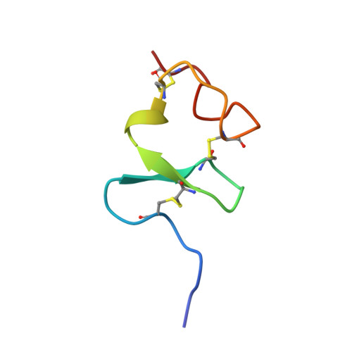
The receptors for the peptide hormones relaxin and insulin-like peptide 3 (INSL3) are the leucine-rich repeat-containing G-protein-coupled receptors LGR7 and LGR8 recently renamed as the relaxin family peptide (RXFP) receptors, RXFP1 and RXFP2, respectively. These receptors differ from other LGRs by the addition of an N-terminal low density lipoprotein receptor class A (LDLa) module and are the only human G-protein-coupled receptors to contain such a domain. Recently it was shown that the LDLa module of the RXFP1 and RXFP2 receptors is essential for ligand-stimulated cAMP signaling. The mechanism by which the LDLa module modulates receptor signaling is unknown; however, it represents a unique paradigm in understanding G-protein-coupled receptor signaling. Here we present the structure of the RXFP1 receptor LDLa module determined by solution NMR spectroscopy. The structure is similar to other LDLa modules but shows small differences in side chain orientations and inter-residue packing. Interchange of the module with the second ligand binding domain of the LDL receptor, LB2, results in a receptor that binds relaxin with full affinity but is unable to signal. Furthermore, we demonstrate via structural studies on mutated LDLa modules and functional studies on mutated full-length receptors that a hydrophobic surface within the N-terminal region of the module is essential for activation of RXFP1 receptor signal in response to relaxin stimulation. This study has highlighted the necessity to understand the structural effects of single amino acid mutations on the LDLa module to fully interpret the effects of these mutations on receptor activity.
Organizational Affiliation:
Department of Biochemistry and Molecular Biology, Bio21 Molecular Science and Biotechnology Institute, University of Melbourne, Victoria 3010, Australia.