2JLM

Structure of a Putative Acetyltransferase (ACIAD1637) from Acinetobacter baylyi ADP1


Experimental Data Snapshot

  • Method: X-RAY DIFFRACTION
  • Resolution: 2.35 Å
  • R-Value Free: 0.238 
  • R-Value Work: 0.182 

wwPDB Validation   3D Report Full Report


This is version 1.5 of the entry. See complete history


Literature

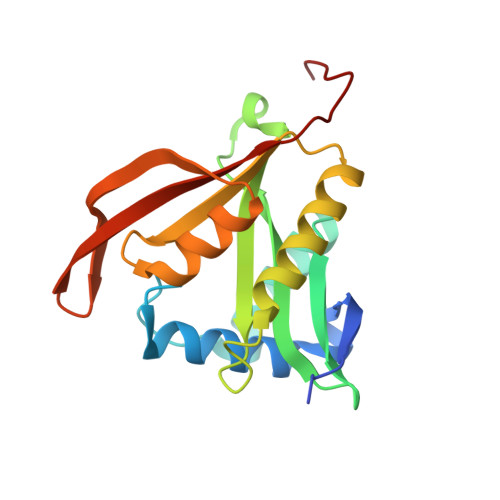
Structure and Substrate Specificity of Acetyltransferase Aciad1637 from Acinetobacter Baylyi Adp1.

Davies, A.M.Tata, R.Snape, A.Sutton, B.J.Brown, P.R.

(2009) Biochimie 91: 484

  • DOI: https://doi.org/10.1016/j.biochi.2008.12.003
  • Primary Citation of Related Structures:  
    2JLM

  • PubMed Abstract: 

    Gene ACIAD1637 from Acinetobacter baylyi ADP1 encodes a 182 amino acid putative antibiotic resistance protein. The structure of this protein (termed acepita) has been solved in space group P(2) to 2.35 A resolution. Acepita belongs to the GCN5-related N-acetyltransferase (GNAT) family, and contains the four sequence motifs conserved among family members. The structure of acepita is compared with that of pita, its homologue from Pseudomonas aeruginosa. Acepita has a similar substrate profile to pita and performs a similar function.


  • Organizational Affiliation

    King's College London, Randall Division of Cell and Molecular Biophysics, London, SE1 1UL, UK.


Macromolecules
Find similar proteins by:  (by identity cutoff)  |  3D Structure
Entity ID: 1
MoleculeChains Sequence LengthOrganismDetailsImage
PUTATIVE PHOSPHINOTHRICIN N-ACETYLTRANSFERASE
A, B, C, D, E
A, B, C, D, E, F
182Acinetobacter baylyiMutation(s): 0 
UniProt
Find proteins for Q6FBS8 (Acinetobacter baylyi (strain ATCC 33305 / BD413 / ADP1))
Explore Q6FBS8 
Go to UniProtKB:  Q6FBS8
Entity Groups  
Sequence Clusters30% Identity50% Identity70% Identity90% Identity95% Identity100% Identity
UniProt GroupQ6FBS8
Sequence Annotations
Expand
  • Reference Sequence
Small Molecules
Ligands 4 Unique
IDChains Name / Formula / InChI Key2D Diagram3D Interactions
PEG
Query on PEG

Download Ideal Coordinates CCD File 
CA [auth E]
HA [auth F]
I [auth A]
K [auth B]
S [auth C]
CA [auth E],
HA [auth F],
I [auth A],
K [auth B],
S [auth C],
V [auth D]
DI(HYDROXYETHYL)ETHER
C4 H10 O3
MTHSVFCYNBDYFN-UHFFFAOYSA-N
ACT
Query on ACT

Download Ideal Coordinates CCD File 
EA [auth F]
G [auth A]
H [auth A]
J [auth B]
L [auth C]
EA [auth F],
G [auth A],
H [auth A],
J [auth B],
L [auth C],
M [auth C],
N [auth C],
O [auth C],
P [auth C],
Q [auth C],
U [auth D],
W [auth E],
X [auth E],
Y [auth E],
Z [auth E]
ACETATE ION
C2 H3 O2
QTBSBXVTEAMEQO-UHFFFAOYSA-M
AZI
Query on AZI

Download Ideal Coordinates CCD File 
AA [auth E],
BA [auth E],
FA [auth F],
GA [auth F],
R [auth C]
AZIDE ION
N3
IVRMZWNICZWHMI-UHFFFAOYSA-N
NA
Query on NA

Download Ideal Coordinates CCD File 
DA [auth E],
T [auth C]
SODIUM ION
Na
FKNQFGJONOIPTF-UHFFFAOYSA-N
Experimental Data & Validation

Experimental Data

  • Method: X-RAY DIFFRACTION
  • Resolution: 2.35 Å
  • R-Value Free: 0.238 
  • R-Value Work: 0.182 
  • Space Group: P 32
Unit Cell:
Length ( Å )Angle ( ˚ )
a = 78.41α = 90
b = 78.41β = 90
c = 197.76γ = 120
Software Package:
Software NamePurpose
PHENIXrefinement
MOSFLMdata reduction
SCALAdata scaling
MOLREPphasing

Structure Validation

View Full Validation Report


Entry History 

Deposition Data

Revision History  (Full details and data files)

  • Version 1.0: 2009-01-20
    Type: Initial release
  • Version 1.1: 2011-05-07
    Changes: Version format compliance
  • Version 1.2: 2011-07-13
    Changes: Version format compliance
  • Version 1.3: 2019-05-08
    Changes: Data collection, Experimental preparation, Other
  • Version 1.4: 2019-06-12
    Changes: Data collection, Database references
  • Version 1.5: 2023-12-13
    Changes: Data collection, Database references, Derived calculations, Other, Refinement description