2JKT

AP2 CLATHRIN ADAPTOR CORE with CD4 Dileucine peptide RM(phosphoS) EIKRLLSE Q to E mutant


Experimental Data Snapshot

  • Method: X-RAY DIFFRACTION
  • Resolution: 3.40 Å
  • R-Value Free: 0.256 
  • R-Value Work: 0.202 
  • R-Value Observed: 0.205 

wwPDB Validation   3D Report Full Report


This is version 1.2 of the entry. See complete history


Literature

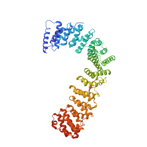
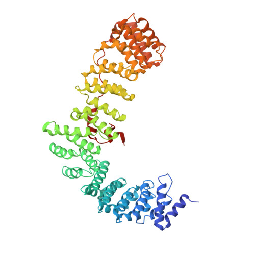
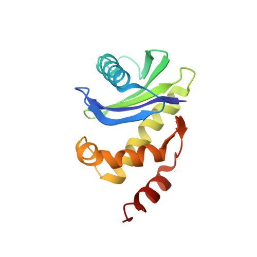
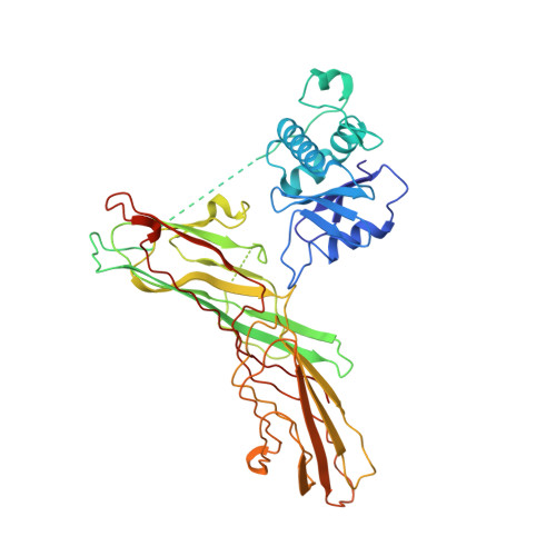
A Structural Explanation for the Binding of Endocytic Dileucine Motifs by the Ap2 Complex.

Kelly, B.T.Mccoy, A.J.Spaete, K.Miller, S.E.Evans, P.R.Hoening, S.Owen, D.J.

(2008) Nature 456: 976

  • DOI: https://doi.org/10.1038/nature07422
  • Primary Citation of Related Structures:  
    2JKR, 2JKT

  • PubMed Abstract: 

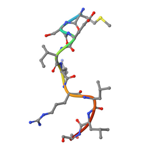
    Most transmembrane proteins are selected as transport vesicle cargo through the recognition of short, linear amino acid motifs in their cytoplasmic portions by vesicle coat proteins. In the case of clathrin-coated vesicles (CCVs) the motifs are recognised by clathrin adaptors. The AP2 adaptor complex (subunits α,β2,μ2,σ2) recognises both major endocytic motifs: YxxΦ motifs and [DE]xxxL[LI] acidic dileucine motifs. Here we describe the binding of AP2 to the endocytic dileucine motif from CD4 . The major recognition events are the two leucine residues binding in hydrophobic pockets on σ2. The hydrophilic residue four residues upstream from the first leucine sits on a positively charged patch made from residues on σ2 and α subunits. Mutations in key residues inhibit the binding of AP2 to ‘acidic dileucine’ motifs displayed in liposomes containing PtdIns4,5P 2 , but do not affect binding to YxxΦ motifs via μ2. In the ‘inactive’ AP2 core structure , both motif binding sites are blocked by different parts of the β2 subunit. To allow a dileucine motif to bind, the β2 N-terminus is displaced and becomes disordered; however, in this structure the YxxΦ binding site on μ2 remains blocked.


  • Organizational Affiliation

    Cambridge Institute for Medical Research and Department of Clinical Biochemistry, University of Cambridge, Addenbrooke's Hospital, Hills Road, Cambridge. CB2 0XY, UK.


Macromolecules
Find similar proteins by:  (by identity cutoff)  |  3D Structure
Entity ID: 1
MoleculeChains Sequence LengthOrganismDetailsImage
AP-2 COMPLEX SUBUNIT ALPHA-2A,
E [auth L]
623Mus musculusMutation(s): 0 
UniProt & NIH Common Fund Data Resources
Find proteins for P17427 (Mus musculus)
Explore P17427 
Go to UniProtKB:  P17427
IMPC:  MGI:101920
Entity Groups  
Sequence Clusters30% Identity50% Identity70% Identity90% Identity95% Identity100% Identity
UniProt GroupP17427
Sequence Annotations
Expand
  • Reference Sequence
Find similar proteins by:  (by identity cutoff)  |  3D Structure
Entity ID: 2
MoleculeChains Sequence LengthOrganismDetailsImage
AP-2 COMPLEX SUBUNIT BETA-1B,
C [auth E]
591Homo sapiensMutation(s): 0 
UniProt & NIH Common Fund Data Resources
Find proteins for P63010 (Homo sapiens)
Explore P63010 
Go to UniProtKB:  P63010
PHAROS:  P63010
GTEx:  ENSG00000006125 
Entity Groups  
Sequence Clusters30% Identity50% Identity70% Identity90% Identity95% Identity100% Identity
UniProt GroupP63010
Sequence Annotations
Expand
  • Reference Sequence
Find similar proteins by:  (by identity cutoff)  |  3D Structure
Entity ID: 3
MoleculeChains Sequence LengthOrganismDetailsImage
AP-2 COMPLEX SUBUNIT SIGMA-1D [auth I],
I [auth S]
142Mus musculusMutation(s): 0 
UniProt & NIH Common Fund Data Resources
Find proteins for P62743 (Mus musculus)
Explore P62743 
Go to UniProtKB:  P62743
IMPC:  MGI:2141861
Entity Groups  
Sequence Clusters30% Identity50% Identity70% Identity90% Identity95% Identity100% Identity
UniProt GroupP62743
Sequence Annotations
Expand
  • Reference Sequence
Find similar proteins by:  (by identity cutoff)  |  3D Structure
Entity ID: 4
MoleculeChains Sequence LengthOrganismDetailsImage
AP-2 COMPLEX SUBUNIT MU-1F [auth M],
J [auth U]
435Rattus norvegicusMutation(s): 0 
UniProt
Find proteins for P84092 (Rattus norvegicus)
Explore P84092 
Go to UniProtKB:  P84092
Entity Groups  
Sequence Clusters30% Identity50% Identity70% Identity90% Identity95% Identity100% Identity
UniProt GroupP84092
Sequence Annotations
Expand
  • Reference Sequence

Find similar proteins by:  Sequence   |   3D Structure  

Entity ID: 5
MoleculeChains Sequence LengthOrganismDetailsImage
CD4 PEPTIDEG [auth P],
H [auth Q]
11Homo sapiensMutation(s): 1 
UniProt & NIH Common Fund Data Resources
Find proteins for P01730 (Homo sapiens)
Explore P01730 
Go to UniProtKB:  P01730
PHAROS:  P01730
GTEx:  ENSG00000010610 
Entity Groups  
Sequence Clusters30% Identity50% Identity70% Identity90% Identity95% Identity100% Identity
UniProt GroupP01730
Sequence Annotations
Expand
  • Reference Sequence
Small Molecules
Ligands 1 Unique
IDChains Name / Formula / InChI Key2D Diagram3D Interactions
SO4
Query on SO4

Download Ideal Coordinates CCD File 
AA [auth L]
BA [auth L]
CA [auth L]
DA [auth L]
EA [auth L]
AA [auth L],
BA [auth L],
CA [auth L],
DA [auth L],
EA [auth L],
FA [auth L],
GA [auth L],
HA [auth L],
IA [auth M],
JA [auth M],
K [auth A],
KA [auth M],
L [auth A],
LA [auth U],
M [auth A],
MA [auth U],
N [auth A],
NA [auth U],
O [auth A],
OA [auth U],
P [auth A],
Q [auth A],
R [auth B],
S [auth B],
T [auth B],
U [auth B],
V [auth B],
W [auth E],
X [auth E],
Y [auth E],
Z [auth E]
SULFATE ION
O4 S
QAOWNCQODCNURD-UHFFFAOYSA-L
Modified Residues  1 Unique
IDChains TypeFormula2D DiagramParent
SEP
Query on SEP
G [auth P],
H [auth Q]
L-PEPTIDE LINKINGC3 H8 N O6 PSER
Experimental Data & Validation

Experimental Data

  • Method: X-RAY DIFFRACTION
  • Resolution: 3.40 Å
  • R-Value Free: 0.256 
  • R-Value Work: 0.202 
  • R-Value Observed: 0.205 
  • Space Group: P 43 21 2
Unit Cell:
Length ( Å )Angle ( ˚ )
a = 171.2α = 90
b = 171.2β = 90
c = 324.27γ = 90
Software Package:
Software NamePurpose
PHENIXrefinement
MOSFLMdata reduction
SCALAdata scaling
PHENIXphasing

Structure Validation

View Full Validation Report


Entry History 

Deposition Data

Revision History  (Full details and data files)

  • Version 1.0: 2008-10-28
    Type: Initial release
  • Version 1.1: 2012-06-20
    Changes: Database references, Derived calculations, Non-polymer description, Other, Refinement description, Version format compliance
  • Version 1.2: 2023-12-13
    Changes: Data collection, Database references, Derived calculations, Other, Refinement description