2JKN

DraE Adhesin in complex with Chloramphenicol Succinate (trigonal form)


Experimental Data Snapshot

  • Method: X-RAY DIFFRACTION
  • Resolution: 1.90 Å
  • R-Value Free: 0.212 
  • R-Value Work: 0.191 
  • R-Value Observed: 0.192 

wwPDB Validation   3D Report Full Report


Ligand Structure Quality Assessment 


This is version 1.3 of the entry. See complete history


Literature

A structural study of the interaction between the Dr haemagglutinin DraE and derivatives of chloramphenicol.

Pettigrew, D.M.Roversi, P.Davies, S.G.Russell, A.J.Lea, S.M.

(2009) Acta Crystallogr D Biol Crystallogr 65: 513-522

  • DOI: https://doi.org/10.1107/S0907444909005113
  • Primary Citation of Related Structures:  
    2JKJ, 2JKL, 2JKN, 2W5P

  • PubMed Abstract: 

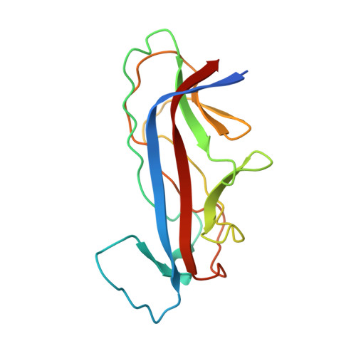
    Dr adhesins are expressed on the surface of uropathogenic and diffusely adherent strains of Escherichia coli. The major adhesin subunit (DraE/AfaE) of these organelles mediates attachment of the bacterium to the surface of the host cell and possibly intracellular invasion through its recognition of the complement regulator decay-accelerating factor (DAF) and/or members of the carcinoembryonic antigen (CEA) family. The adhesin subunit of the Dr haemagglutinin, a Dr-family member, additionally binds type IV collagen and is inhibited in all its receptor interactions by the antibiotic chloramphenicol (CLM). In this study, previous structural work is built upon by reporting the X-ray structures of DraE bound to two chloramphenicol derivatives: chloramphenicol succinate (CLS) and bromamphenicol (BRM). The CLS structure demonstrates that acylation of the 3-hydroxyl group of CLM with succinyl does not significantly perturb the mode of binding, while the BRM structure implies that the binding pocket is able to accommodate bulkier substituents on the N-acyl group. It is concluded that modifications of the 3-hydroxyl group would generate a potent Dr haemagglutinin inhibitor that would not cause the toxic side effects that are associated with the normal bacteriostatic activity of CLM.


  • Organizational Affiliation

    Sir William Dunn School of Pathology, University of Oxford, Oxford OX1 3RE, England.


Macromolecules
Find similar proteins by:  (by identity cutoff)  |  3D Structure
Entity ID: 1
MoleculeChains Sequence LengthOrganismDetailsImage
DR HEMAGGLUTININ STRUCTURAL SUBUNIT
A, B, C, D, E
A, B, C, D, E, F
149Escherichia coliMutation(s): 0 
UniProt
Find proteins for P24093 (Escherichia coli)
Explore P24093 
Go to UniProtKB:  P24093
Entity Groups  
Sequence Clusters30% Identity50% Identity70% Identity90% Identity95% Identity100% Identity
UniProt GroupP24093
Sequence Annotations
Expand
  • Reference Sequence
Small Molecules
Ligands 3 Unique
IDChains Name / Formula / InChI Key2D Diagram3D Interactions
CL8
Query on CL8

Download Ideal Coordinates CCD File 
CA [auth E]
HA [auth F]
I [auth A]
N [auth B]
S [auth C]
CA [auth E],
HA [auth F],
I [auth A],
N [auth B],
S [auth C],
X [auth D]
CHLORAMPHENICOL SUCCINATE
C15 H16 Cl2 N2 O8
LIRCDOVJWUGTMW-ZWNOBZJWSA-N
SO4
Query on SO4

Download Ideal Coordinates CCD File 
AA [auth E]
BA [auth E]
FA [auth F]
G [auth A]
GA [auth F]
AA [auth E],
BA [auth E],
FA [auth F],
G [auth A],
GA [auth F],
H [auth A],
L [auth B],
M [auth B],
Q [auth C],
R [auth C],
V [auth D],
W [auth D]
SULFATE ION
O4 S
QAOWNCQODCNURD-UHFFFAOYSA-L
EDO
Query on EDO

Download Ideal Coordinates CCD File 
DA [auth E]
EA [auth E]
IA [auth F]
J [auth A]
JA [auth F]
DA [auth E],
EA [auth E],
IA [auth F],
J [auth A],
JA [auth F],
K [auth A],
O [auth B],
P [auth B],
T [auth C],
U [auth C],
Y [auth D],
Z [auth D]
1,2-ETHANEDIOL
C2 H6 O2
LYCAIKOWRPUZTN-UHFFFAOYSA-N
Experimental Data & Validation

Experimental Data

  • Method: X-RAY DIFFRACTION
  • Resolution: 1.90 Å
  • R-Value Free: 0.212 
  • R-Value Work: 0.191 
  • R-Value Observed: 0.192 
  • Space Group: P 3
Unit Cell:
Length ( Å )Angle ( ˚ )
a = 119.3α = 90
b = 119.3β = 90
c = 57.7γ = 120
Software Package:
Software NamePurpose
BUSTER-TNTrefinement
MOSFLMdata reduction
SCALAdata scaling
MOLREPphasing

Structure Validation

View Full Validation Report


Ligand Structure Quality Assessment 


Entry History 

Deposition Data

Revision History  (Full details and data files)

  • Version 1.0: 2008-12-16
    Type: Initial release
  • Version 1.1: 2012-09-12
    Changes: Database references, Derived calculations, Non-polymer description, Other, Refinement description, Structure summary
  • Version 1.2: 2018-06-06
    Changes: Data collection, Database references, Derived calculations
  • Version 1.3: 2023-12-13
    Changes: Data collection, Database references, Derived calculations, Other, Refinement description