2JK9

The structure of splA-ryanodine receptor domain and SOCS box containing 1 in complex with a PAR-4 peptide


Experimental Data Snapshot

  • Method: X-RAY DIFFRACTION
  • Resolution: 1.79 Å
  • R-Value Free: 0.240 
  • R-Value Work: 0.192 
  • R-Value Observed: 0.194 

wwPDB Validation   3D Report Full Report


This is version 1.3 of the entry. See complete history


Literature

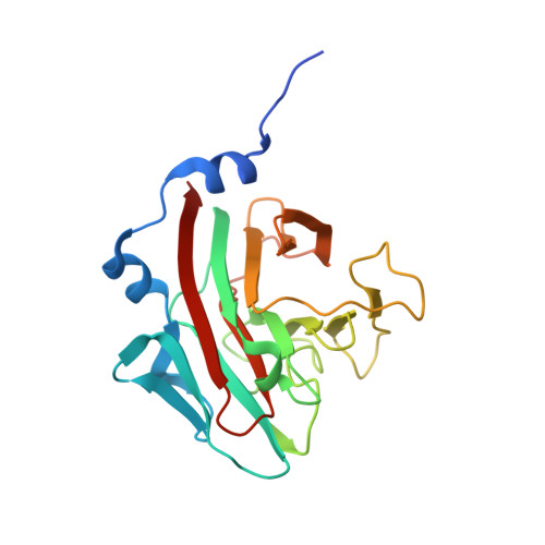
Structural Basis for Par-4 Recognition by the Spry Domain-and Socs Box-Containing Proteins Spsb1, Spsb2, and Spsb4.

Filippakopoulos, P.Low, A.Sharpe, T.D.Uppenberg, J.Yao, S.Kuang, Z.Savitsky, P.Lewis, R.S.Nicholson, S.E.Norton, R.S.Bullock, A.

(2010) J Mol Biol 401: 389

  • DOI: https://doi.org/10.1016/j.jmb.2010.06.017
  • Primary Citation of Related Structures:  
    2JK9, 2V24, 3EMW, 3F2O

  • PubMed Abstract: 

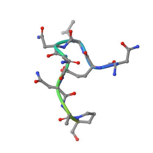
    The mammalian SPRY domain- and SOCS box-containing proteins, SPSB1 to SPSB4, belong to the SOCS box family of E3 ubiquitin ligases. Substrate recognition sites for the SPRY domain are identified only for human Par-4 (ELNNNL) and for the Drosophila orthologue GUSTAVUS binding to the DEAD-box RNA helicase VASA (DINNNN). To further investigate this consensus motif, we determined the crystal structures of SPSB1, SPSB2, and SPSB4, as well as their binding modes and affinities for both Par-4 and VASA. Mutation of each of the three Asn residues in Par-4 abrogated binding to all three SPSB proteins, while changing EL to DI enhanced binding. By comparison to SPSB1 and SPSB4, the more divergent protein SPSB2 showed only weak binding to Par-4 and was hypersensitive to DI substitution. Par-4((59-77)) binding perturbed NMR resonances from a number of SPSB2 residues flanking the ELNNN binding site, including loop D, which binds the EL/DI sequence. Although interactions with the consensus peptide motif were conserved in all structures, flanking sites in SPSB2 were identified as sites of structural change. These structural changes limit high-affinity interactions for SPSB2 to aspartate-containing sequences, whereas SPSB1 and SPSB4 bind strongly to both Par-4 and VASA peptides.


  • Organizational Affiliation

    Structural Genomics Consortium, University of Oxford, Old Road Campus, Roosevelt Drive, Oxford OX3 7DQ, UK.


Macromolecules
Find similar proteins by:  (by identity cutoff)  |  3D Structure
Entity ID: 1
MoleculeChains Sequence LengthOrganismDetailsImage
SPRY DOMAIN-CONTAINING SOCS BOX PROTEIN 1212Homo sapiensMutation(s): 0 
UniProt & NIH Common Fund Data Resources
Find proteins for Q96BD6 (Homo sapiens)
Explore Q96BD6 
Go to UniProtKB:  Q96BD6
PHAROS:  Q96BD6
GTEx:  ENSG00000171621 
Entity Groups  
Sequence Clusters30% Identity50% Identity70% Identity90% Identity95% Identity100% Identity
UniProt GroupQ96BD6
Sequence Annotations
Expand
  • Reference Sequence

Find similar proteins by:  Sequence   |   3D Structure  

Entity ID: 2
MoleculeChains Sequence LengthOrganismDetailsImage
PRKC APOPTOSIS WT1 REGULATOR PROTEIN15Homo sapiensMutation(s): 0 
UniProt & NIH Common Fund Data Resources
Find proteins for Q96IZ0 (Homo sapiens)
Explore Q96IZ0 
Go to UniProtKB:  Q96IZ0
PHAROS:  Q96IZ0
GTEx:  ENSG00000177425 
Entity Groups  
Sequence Clusters30% Identity50% Identity70% Identity90% Identity95% Identity100% Identity
UniProt GroupQ96IZ0
Sequence Annotations
Expand
  • Reference Sequence
Experimental Data & Validation

Experimental Data

  • Method: X-RAY DIFFRACTION
  • Resolution: 1.79 Å
  • R-Value Free: 0.240 
  • R-Value Work: 0.192 
  • R-Value Observed: 0.194 
  • Space Group: P 1 21 1
Unit Cell:
Length ( Å )Angle ( ˚ )
a = 37.523α = 90
b = 82.957β = 104.42
c = 38.54γ = 90
Software Package:
Software NamePurpose
REFMACrefinement
MOSFLMdata reduction
SCALAdata scaling
PHASERphasing

Structure Validation

View Full Validation Report


Entry History 

Revision History  (Full details and data files)

  • Version 1.0: 2008-09-16
    Type: Initial release
  • Version 1.1: 2011-07-13
    Changes: Advisory, Version format compliance
  • Version 1.2: 2012-12-05
    Changes: Structure summary
  • Version 1.3: 2023-12-13
    Changes: Data collection, Database references, Other, Refinement description