The Crystal Structure of Pyrococcus Abyssi tRNA (Uracil-54, C5)-Methyltransferase Provides Insights Into its tRNA Specificity.
Walbott, H., Leulliot, N., Grosjean, H., Golinelli-Pimpaneau, B.(2008) Nucleic Acids Res 36: 4929
- PubMed: 18653523
- DOI: https://doi.org/10.1093/nar/gkn437
- Primary Citation of Related Structures:
2JJQ, 2VS1 - PubMed Abstract:
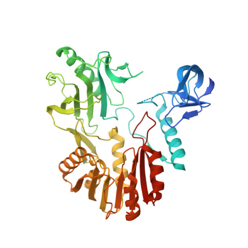
The 5-methyluridine is invariably found at position 54 in the TPsiC loop of tRNAs of most organisms. In Pyrococcus abyssi, its formation is catalyzed by the S-adenosyl-l-methionine-dependent tRNA (uracil-54, C5)-methyltransferase ((Pab)TrmU54), an enzyme that emerged through an ancient horizontal transfer of an RNA (uracil, C5)-methyltransferase-like gene from bacteria to archaea. The crystal structure of (Pab)TrmU54 in complex with S-adenosyl-l-homocysteine at 1.9 A resolution shows the protein organized into three domains like Escherichia coli RumA, which catalyzes the same reaction at position 1939 of 23S rRNA. A positively charged groove at the interface between the three domains probably locates part of the tRNA-binding site of (Pab)TrmU54. We show that a mini-tRNA lacking both the D and anticodon stem-loops is recognized by (Pab)TrmU54. These results were used to model yeast tRNA(Asp) in the (Pab)TrmU54 structure to get further insights into the different RNA specificities of RumA and (Pab)TrmU54. Interestingly, the presence of two flexible loops in the central domain, unique to (Pab)TrmU54, may explain the different substrate selectivities of both enzymes. We also predict that a large TPsiC loop conformational change has to occur for the flipping of the target uridine into the (Pab)TrmU54 active site during catalysis.
Organizational Affiliation:
Enzymology and Structural Biochemistry Laboratory, CNRS, 1 avenue de la Terrasse, 91198 Gif-sur-Yvette, France.