2JJ7

Crystal structure of the HlyIIR mutant protein with residues 170-185 substituted by alanine


Experimental Data Snapshot

  • Method: X-RAY DIFFRACTION
  • Resolution: 2.10 Å
  • R-Value Free: 0.237 
  • R-Value Work: 0.179 
  • R-Value Observed: 0.181 

wwPDB Validation   3D Report Full Report


This is version 1.4 of the entry. See complete history


Literature

[Contraction of the disordered loop located within C-terminal domain of the transcriptional regulator HlyIIR causes its structural rearrangement].

Kovalevskii, O.V.Antson, A.A.Solonin, A.S.

(2009) Mol Biol (Mosk) 43: 126-135

  • Primary Citation of Related Structures:  
    2JJ7

  • PubMed Abstract: 

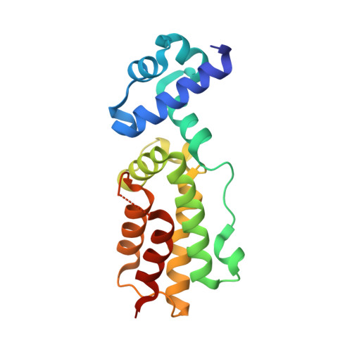
    HlyIIR is a negative transcriptional regulator of the hemolysin II gene from Bacillus cereus . A disordered region (amino acid residues 170-185) localized within the C-terminal domain near the dimerization interface was found in the recently determined HlyIIR X-ray structure. To clarify the effect of this region on HlyIIR properties and potential improvement of the diffraction quality of its crystals, we constructed a HlyIIR mutant with a single alanine residue substituting for the overall disordered region. According to biochemical analysis, the mutant protein still formed a dimer but lost its DNA-binding activity. Its crystals displayed better diffraction quality as compared with the native protein. The mutant structure was determined by X-ray analysis with a resolution of 2.1 Å. However, the mutant protein formed an alternative dimer differing from the wild-type dimer, as its subunits were rotated by 160. The conformation of individual subunits also partially changed. As this considerable remodeling in the mutant protein structure resulted from the conformational changes in the segment Pro161-Ser169, we concluded that this segment was important for maintaining the native HlyIIR structure.


Macromolecules
Find similar proteins by:  (by identity cutoff)  |  3D Structure
Entity ID: 1
MoleculeChains Sequence LengthOrganismDetailsImage
HEMOLYSIN II REGULATORY PROTEIN
A, B
186Bacillus cereusMutation(s): 0 
UniProt
Find proteins for Q7X506 (Bacillus cereus)
Explore Q7X506 
Go to UniProtKB:  Q7X506
Entity Groups  
Sequence Clusters30% Identity50% Identity70% Identity90% Identity95% Identity100% Identity
UniProt GroupQ7X506
Sequence Annotations
Expand
  • Reference Sequence
Experimental Data & Validation

Experimental Data

  • Method: X-RAY DIFFRACTION
  • Resolution: 2.10 Å
  • R-Value Free: 0.237 
  • R-Value Work: 0.179 
  • R-Value Observed: 0.181 
  • Space Group: P 21 21 2
Unit Cell:
Length ( Å )Angle ( ˚ )
a = 66.831α = 90
b = 120.414β = 90
c = 55.178γ = 90
Software Package:
Software NamePurpose
REFMACrefinement
DENZOdata reduction
SCALEPACKdata scaling
MOLREPphasing

Structure Validation

View Full Validation Report


Entry History 

Deposition Data

Revision History  (Full details and data files)

  • Version 1.0: 2008-04-08
    Type: Initial release
  • Version 1.1: 2011-07-13
    Changes: Advisory, Refinement description, Version format compliance
  • Version 1.2: 2011-12-28
    Changes: Other
  • Version 1.3: 2019-10-09
    Changes: Data collection, Database references, Other
  • Version 1.4: 2023-12-13
    Changes: Data collection, Database references, Refinement description