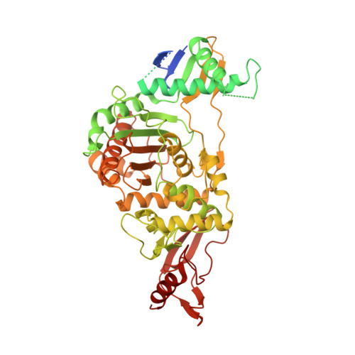
2JCH

Structural and mechanistic basis of penicillin binding protein inhibition by lactivicins


Experimental Data Snapshot

  • Method: X-RAY DIFFRACTION
  • Resolution: 2.40 Å
  • R-Value Free: 0.262 
  • R-Value Work: 0.203 
  • R-Value Observed: 0.206 

wwPDB Validation   3D Report Full Report


Ligand Structure Quality Assessment 


This is version 1.3 of the entry. See complete history


Literature

Structural and Mechanistic Basis of Penicillin-Binding Protein Inhibition by Lactivicins

Macheboeuf, P.Fisher, D.S.Brown, T.J.Zervosen, A.Luxen, A.Joris, B.Dessen, A.Schofield, C.J.

(2007) Nat Chem Biol 3: 565

  • DOI: https://doi.org/10.1038/nchembio.2007.21
  • Primary Citation of Related Structures:  
    2JCH, 2JE5

  • PubMed Abstract: 

    Beta-lactam antibiotics, including penicillins and cephalosporins, inhibit penicillin-binding proteins (PBPs), which are essential for bacterial cell wall biogenesis. Pathogenic bacteria have evolved efficient antibiotic resistance mechanisms that, in Gram-positive bacteria, include mutations to PBPs that enable them to avoid beta-lactam inhibition. Lactivicin (LTV; 1) contains separate cycloserine and gamma-lactone rings and is the only known natural PBP inhibitor that does not contain a beta-lactam. Here we show that LTV and a more potent analog, phenoxyacetyl-LTV (PLTV; 2), are active against clinically isolated, penicillin-resistant Streptococcus pneumoniae strains. Crystallographic analyses of S. pneumoniae PBP1b reveal that LTV and PLTV inhibition involves opening of both monocyclic cycloserine and gamma-lactone rings. In PBP1b complexes, the ring-derived atoms from LTV and PLTV show a notable structural convergence with those derived from a complexed cephalosporin (cefotaxime; 3). The structures imply that derivatives of LTV will be useful in the search for new antibiotics with activity against beta-lactam-resistant bacteria.


  • Organizational Affiliation

    Institut de Biologie Structurale Jean-Pierre Ebel Commissariat à l'énergie atomique - Centre National de La Recherche Scientifique - Université Joseph Fourier, 41 rue Jules Horowitz, F-38027 Grenoble, France.


Macromolecules
Find similar proteins by:  (by identity cutoff)  |  3D Structure
Entity ID: 1
MoleculeChains Sequence LengthOrganismDetailsImage
PENICILLIN-BINDING PROTEIN 1B720Streptococcus pneumoniae R6Mutation(s): 7 
UniProt
Find proteins for O70038 (Streptococcus pneumoniae)
Explore O70038 
Go to UniProtKB:  O70038
Entity Groups  
Sequence Clusters30% Identity50% Identity70% Identity90% Identity95% Identity100% Identity
UniProt GroupO70038
Sequence Annotations
Expand
  • Reference Sequence
Experimental Data & Validation

Experimental Data

  • Method: X-RAY DIFFRACTION
  • Resolution: 2.40 Å
  • R-Value Free: 0.262 
  • R-Value Work: 0.203 
  • R-Value Observed: 0.206 
  • Space Group: C 2 2 21
Unit Cell:
Length ( Å )Angle ( ˚ )
a = 105.84α = 90
b = 143.58β = 90
c = 97.44γ = 90
Software Package:
Software NamePurpose
REFMACrefinement
XDSdata reduction
XDSdata scaling
AMoREphasing

Structure Validation

View Full Validation Report


Ligand Structure Quality Assessment 


Entry History 

Deposition Data

Revision History  (Full details and data files)

  • Version 1.0: 2007-08-14
    Type: Initial release
  • Version 1.1: 2011-05-08
    Changes: Version format compliance
  • Version 1.2: 2011-07-13
    Changes: Version format compliance
  • Version 1.3: 2023-12-13
    Changes: Data collection, Database references, Derived calculations, Other, Refinement description