


Crystal Structure of Proteasome Beta Subunit Prcb from Mycobacterium Tuberculosis
Oberschall, A., Strizhov, N., Bartunik, H.D.To be published.
Experimental Data Snapshot
wwPDB Validation 3D Report Full Report

Entity ID: 1 | |||||
|---|---|---|---|---|---|
| Molecule | Chains | Sequence Length | Organism | Details | Image |
| PROTEASOME | 291 | Mycobacterium tuberculosis H37Rv | Mutation(s): 0 EC: 3.4.25.1 | 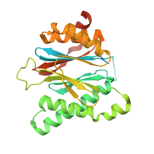 | |
UniProt | |||||
Find proteins for P9WHT9 (Mycobacterium tuberculosis (strain ATCC 25618 / H37Rv)) Explore P9WHT9 Go to UniProtKB: P9WHT9 | |||||
Entity Groups | |||||
| Sequence Clusters | 30% Identity50% Identity70% Identity90% Identity95% Identity100% Identity | ||||
| UniProt Group | P9WHT9 | ||||
Sequence AnnotationsExpand | |||||
| |||||
| Length ( Å ) | Angle ( ˚ ) |
|---|---|
| a = 40.504 | α = 90 |
| b = 40.473 | β = 97.71 |
| c = 59.937 | γ = 90 |
| Software Name | Purpose |
|---|---|
| REFMAC | refinement |
| DENZO | data reduction |
| SCALEPACK | data scaling |
| MOLREP | phasing |
RCSB PDB (citation) is hosted by
RCSB PDB is a member of the

