2J9I

Lengsin is a survivor of an ancient family of class I glutamine synthetases in eukaryotes that has undergone evolutionary re- engineering for a tissue-specific role in the vertebrate eye lens.


Experimental Data Snapshot

  • Method: ELECTRON MICROSCOPY
  • Resolution: 17.0 Å
  • Aggregation State: PARTICLE 
  • Reconstruction Method: SINGLE PARTICLE 

wwPDB Validation   3D Report Full Report


This is version 1.3 of the entry. See complete history


Literature

Lengsin is a Survivor of an Ancient Family of Class I Glutamine Synthetases Re-Engineered by Evolution for a Role in the Vertebrate Lens.

Wyatt, K.White, H.E.Wang, L.Bateman, O.A.Slingsby, C.Orlova, E.V.Wistow, G.

(2006) Structure 14: 1823

  • DOI: https://doi.org/10.1016/j.str.2006.10.008
  • Primary Citation of Related Structures:  
    2J9I

  • PubMed Abstract: 

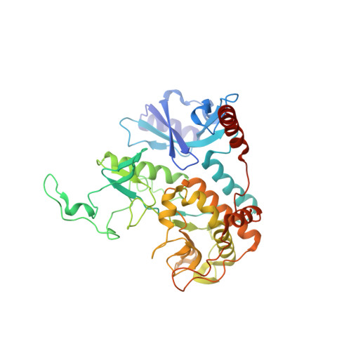
    Lengsin is a major protein of the vertebrate eye lens. It belongs to the hitherto purely prokaryotic GS I branch of the glutamine synthetase (GS) superfamily, but has no enzyme activity. Like the taxon-specific crystallins, Lengsin is the result of the recruitment of an ancient enzyme to a noncatalytic role in the vertebrate lens. Cryo-EM and modeling studies of Lengsin show a dodecamer structure with important similarities and differences with prokaryotic GS I structures. GS homology regions of Lengsin are well conserved, but the N-terminal domain shows evidence of dynamic evolutionary changes. Compared with birds and fish, most mammals have an additional exon corresponding to part of the N-terminal domain; however, in human, this is a nonfunctional pseudoexon. Genes related to Lengsin are also present in the sea urchin, suggesting that this branch of the GS I family, supplanted by GS II enzymes in vertebrates, has an ancient role in metazoans.


  • Organizational Affiliation

    Section on Molecular Structure and Functional Genomics, National Eye Institute, National Institutes of Health, Bethesda, Maryland 20892, USA.


Macromolecules
Find similar proteins by:  (by identity cutoff)  |  3D Structure
Entity ID: 1
MoleculeChains Sequence LengthOrganismDetailsImage
GLUTAMATE-AMMONIA LIGASE DOMAIN-CONTAINING PROTEIN 1
A, B, C, D, E
A, B, C, D, E, F, G, H, I, J, K, L
421Mus musculusMutation(s): 0 
UniProt
Find proteins for Q8CIX8 (Mus musculus)
Explore Q8CIX8 
Go to UniProtKB:  Q8CIX8
Entity Groups  
Sequence Clusters30% Identity50% Identity70% Identity90% Identity95% Identity100% Identity
UniProt GroupQ8CIX8
Sequence Annotations
Expand
  • Reference Sequence
Experimental Data & Validation

Experimental Data

  • Method: ELECTRON MICROSCOPY
  • Resolution: 17.0 Å
  • Aggregation State: PARTICLE 
  • Reconstruction Method: SINGLE PARTICLE 
EM Software:
TaskSoftware PackageVersion
RECONSTRUCTIONIMAGIC

Structure Validation

View Full Validation Report


Entry History 

Deposition Data

Revision History  (Full details and data files)

  • Version 1.0: 2006-12-13
    Type: Initial release
  • Version 1.1: 2013-08-07
    Changes: Derived calculations, Other, Refinement description, Source and taxonomy, Version format compliance
  • Version 1.2: 2017-08-23
    Changes: Data collection
  • Version 1.3: 2019-10-23
    Changes: Data collection, Other