2J59

Crystal structure of the ARF1:ARHGAP21-ArfBD complex


Experimental Data Snapshot

  • Method: X-RAY DIFFRACTION
  • Resolution: 2.10 Å
  • R-Value Free: 0.243 
  • R-Value Work: 0.205 
  • R-Value Observed: 0.209 

wwPDB Validation   3D Report Full Report


Ligand Structure Quality Assessment 


This is version 1.3 of the entry. See complete history


Literature

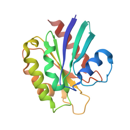
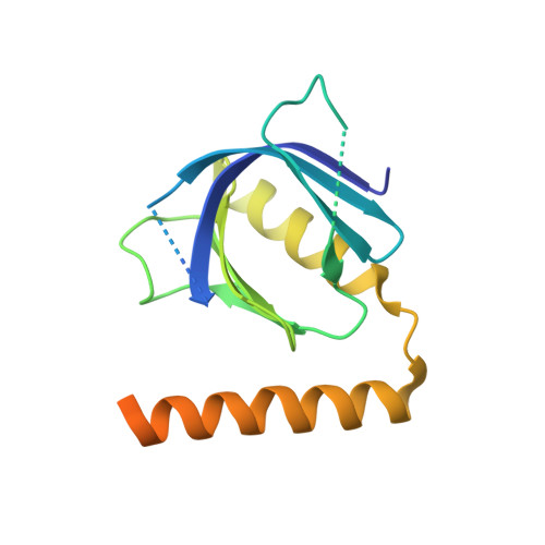
Structural Basis for Arf1-Mediated Recruitment of Arhgap21 to Golgi Membranes.

Menetrey, J.Perderiset, M.Cicolari, J.Dubois, T.El Khatib, N.El Khadali, F.Franco, M.Chavrier, P.Houdusse, A.

(2007) EMBO J 26: 1953

  • DOI: https://doi.org/10.1038/sj.emboj.7601634
  • Primary Citation of Related Structures:  
    2J59

  • PubMed Abstract: 

    ARHGAP21 is a Rho family GTPase-activating protein (RhoGAP) that controls the Arp2/3 complex and F-actin dynamics at the Golgi complex by regulating the activity of the small GTPase Cdc42. ARHGAP21 is recruited to the Golgi by binding to another small GTPase, ARF1. Here, we present the crystal structure of the activated GTP-bound form of ARF1 in a complex with the Arf-binding domain (ArfBD) of ARHGAP21 at 2.1 A resolution. We show that ArfBD comprises a PH domain adjoining a C-terminal alpha helix, and that ARF1 interacts with both of these structural motifs through its switch regions and triggers structural rearrangement of the PH domain. We used site-directed mutagenesis to confirm that both the PH domain and the helical motif are essential for the binding of ArfBD to ARF1 and for its recruitment to the Golgi. Our data demonstrate that two well-known small GTPase-binding motifs, the PH domain and the alpha helical motif, can combine to create a novel mode of binding to Arfs.


  • Organizational Affiliation

    Centre de Recherche, UMR 144, Institut Curie, 26 rue d'Ulm, 75248 Paris Cedex 05, France.


Macromolecules
Find similar proteins by:  (by identity cutoff)  |  3D Structure
Entity ID: 1
MoleculeChains Sequence LengthOrganismDetailsImage
ADP-RIBOSYLATION FACTOR 1
A, B, C, D, E
A, B, C, D, E, F
166Mus musculusMutation(s): 1 
UniProt
Find proteins for P84078 (Mus musculus)
Explore P84078 
Go to UniProtKB:  P84078
Entity Groups  
Sequence Clusters30% Identity50% Identity70% Identity90% Identity95% Identity100% Identity
UniProt GroupP84078
Sequence Annotations
Expand
  • Reference Sequence
Find similar proteins by:  (by identity cutoff)  |  3D Structure
Entity ID: 2
MoleculeChains Sequence LengthOrganismDetailsImage
RHO-GTPASE ACTIVATING PROTEIN 10168Homo sapiensMutation(s): 0 
UniProt & NIH Common Fund Data Resources
Find proteins for Q5T5U3 (Homo sapiens)
Explore Q5T5U3 
Go to UniProtKB:  Q5T5U3
PHAROS:  Q5T5U3
GTEx:  ENSG00000107863 
Entity Groups  
Sequence Clusters30% Identity50% Identity70% Identity90% Identity95% Identity100% Identity
UniProt GroupQ5T5U3
Sequence Annotations
Expand
  • Reference Sequence
Small Molecules
Ligands 5 Unique
IDChains Name / Formula / InChI Key2D Diagram3D Interactions
GTP
Query on GTP

Download Ideal Coordinates CCD File 
AA [auth F]
M [auth A]
P [auth B]
S [auth C]
V [auth D]
AA [auth F],
M [auth A],
P [auth B],
S [auth C],
V [auth D],
X [auth E]
GUANOSINE-5'-TRIPHOSPHATE
C10 H16 N5 O14 P3
XKMLYUALXHKNFT-UUOKFMHZSA-N
SO4
Query on SO4

Download Ideal Coordinates CCD File 
DA [auth M]
EA [auth N]
FA [auth O]
HA [auth P]
IA [auth Q]
DA [auth M],
EA [auth N],
FA [auth O],
HA [auth P],
IA [auth Q],
JA [auth R]
SULFATE ION
O4 S
QAOWNCQODCNURD-UHFFFAOYSA-L
DIO
Query on DIO

Download Ideal Coordinates CCD File 
CA [auth F],
O [auth A],
R [auth B]
1,4-DIETHYLENE DIOXIDE
C4 H8 O2
RYHBNJHYFVUHQT-UHFFFAOYSA-N
EDO
Query on EDO

Download Ideal Coordinates CCD File 
GA [auth O],
U [auth C],
Z [auth E]
1,2-ETHANEDIOL
C2 H6 O2
LYCAIKOWRPUZTN-UHFFFAOYSA-N
MG
Query on MG

Download Ideal Coordinates CCD File 
BA [auth F]
N [auth A]
Q [auth B]
T [auth C]
W [auth D]
BA [auth F],
N [auth A],
Q [auth B],
T [auth C],
W [auth D],
Y [auth E]
MAGNESIUM ION
Mg
JLVVSXFLKOJNIY-UHFFFAOYSA-N
Experimental Data & Validation

Experimental Data

  • Method: X-RAY DIFFRACTION
  • Resolution: 2.10 Å
  • R-Value Free: 0.243 
  • R-Value Work: 0.205 
  • R-Value Observed: 0.209 
  • Space Group: P 1 21 1
Unit Cell:
Length ( Å )Angle ( ˚ )
a = 74.636α = 90
b = 132.136β = 90.01
c = 146.283γ = 90
Software Package:
Software NamePurpose
REFMACrefinement
MOSFLMdata reduction
SCALAdata scaling
PHASERphasing

Structure Validation

View Full Validation Report


Ligand Structure Quality Assessment 


Entry History 

Deposition Data

Revision History  (Full details and data files)

  • Version 1.0: 2007-02-20
    Type: Initial release
  • Version 1.1: 2011-05-08
    Changes: Version format compliance
  • Version 1.2: 2011-07-13
    Changes: Version format compliance
  • Version 1.3: 2023-12-13
    Changes: Data collection, Database references, Derived calculations, Other, Refinement description