2J4K

Crystal structure of uridylate kinase from Sulfolobus solfataricus in complex with UMP to 2.2 Angstrom resolution


Experimental Data Snapshot

  • Method: X-RAY DIFFRACTION
  • Resolution: 2.20 Å
  • R-Value Free: 0.251 
  • R-Value Work: 0.219 
  • R-Value Observed: 0.219 

wwPDB Validation   3D Report Full Report


Ligand Structure Quality Assessment 


This is version 1.4 of the entry. See complete history


Literature

Structural and Enzymatic Investigation of the Sulfolobus Solfataricus Uridylate Kinase Shows Competitive Utp Inhibition and the Lack of GTP Stimulation

Jensen, K.S.Johansson, E.Jensen, K.F.

(2007) Biochemistry 46: 2745

  • DOI: https://doi.org/10.1021/bi0618159
  • Primary Citation of Related Structures:  
    2J4J, 2J4K, 2J4L

  • PubMed Abstract: 

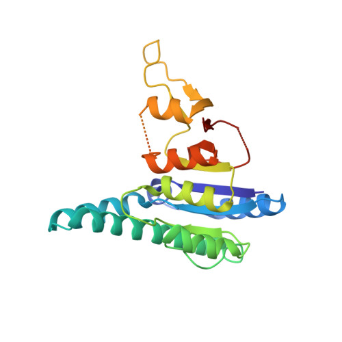
    The pyrH gene encoding uridylate kinase (UMPK) from the extreme thermoacidophilic archaeon Sulfolobus solfataricus was cloned and expressed in Escherichia coli, and the enzyme (SsUMPK) was purified. Size exclusion chromatography and sedimentation experiments showed that the oligomeric state in solution is hexameric. SsUMPK shows maximum catalytic rate at pH 7.0, and variation of pH only influences the turnover number. Catalysis proceeds by a sequential reaction mechanism of random order and depends on a divalent cation. The enzyme exhibits high substrate specificity toward UMP and ATP and is inhibited by UTP, whereas CTP and GTP do not influence activity. UTP binds to the enzyme with a sigmoid binding curve, whereas GTP does not bind. The crystal structure of SsUMPK was determined for three different complexes, a ternary complex with UMP and the nonhydrolyzable ATP analogue beta,gamma-methylene-ATP, a complex with UMP, and a complex with UTP to 2.1, 2.2, and 2.8 A resolution, respectively. One UTP molecule was bound in the acceptor site per subunit, leading to the exclusion of both substrates from the active site. In all cases, SsUMPK crystallized as a hexamer with the main fold shared with other prokaryotic UMPKs. Similar to UMPK from Pyrococcus furiosus, SsUMPK has an active site enclosing loop. This loop was only ordered in one subunit in the ternary complex, which also contained an unusual arrangement of ligands (possibly a dinucleotide) in the active site and an altered orientation of the catalytic residue Arg48 relative to the other five subunits of the hexamer.


  • Organizational Affiliation

    Department of Biological Chemistry, Institute of Molecular Biology, University of Copenhagen, Sølvgade 83H, DK-1307 Copenhagen K, Denmark.


Macromolecules
Find similar proteins by:  (by identity cutoff)  |  3D Structure
Entity ID: 1
MoleculeChains Sequence LengthOrganismDetailsImage
URIDYLATE KINASE
A, B, C, D, E
A, B, C, D, E, F
226Saccharolobus solfataricus P2Mutation(s): 0 
EC: 2.7.4.22
UniProt
Find proteins for Q97ZE2 (Saccharolobus solfataricus (strain ATCC 35092 / DSM 1617 / JCM 11322 / P2))
Explore Q97ZE2 
Go to UniProtKB:  Q97ZE2
Entity Groups  
Sequence Clusters30% Identity50% Identity70% Identity90% Identity95% Identity100% Identity
UniProt GroupQ97ZE2
Sequence Annotations
Expand
  • Reference Sequence
Small Molecules
Ligands 3 Unique
IDChains Name / Formula / InChI Key2D Diagram3D Interactions
U5P
Query on U5P

Download Ideal Coordinates CCD File 
CA [auth F]
G [auth A]
L [auth B]
T [auth C]
X [auth D]
CA [auth F],
G [auth A],
L [auth B],
T [auth C],
X [auth D],
Z [auth E]
URIDINE-5'-MONOPHOSPHATE
C9 H13 N2 O9 P
DJJCXFVJDGTHFX-XVFCMESISA-N
CD
Query on CD

Download Ideal Coordinates CCD File 
AA [auth E]
BA [auth E]
DA [auth F]
EA [auth F]
H [auth A]
AA [auth E],
BA [auth E],
DA [auth F],
EA [auth F],
H [auth A],
I [auth A],
J [auth A],
M [auth B],
N [auth B],
O [auth B],
P [auth B],
Q [auth B],
R [auth B],
S [auth B],
U [auth C],
V [auth C],
W [auth C],
Y [auth D]
CADMIUM ION
Cd
WLZRMCYVCSSEQC-UHFFFAOYSA-N
MG
Query on MG

Download Ideal Coordinates CCD File 
K [auth A]MAGNESIUM ION
Mg
JLVVSXFLKOJNIY-UHFFFAOYSA-N
Binding Affinity Annotations 
IDSourceBinding Affinity
U5P Binding MOAD:  2J4K Kd: 1.40e+4 (nM) from 1 assay(s)
PDBBind:  2J4K Kd: 1.40e+4 (nM) from 1 assay(s)
Experimental Data & Validation

Experimental Data

  • Method: X-RAY DIFFRACTION
  • Resolution: 2.20 Å
  • R-Value Free: 0.251 
  • R-Value Work: 0.219 
  • R-Value Observed: 0.219 
  • Space Group: P 1 21 1
Unit Cell:
Length ( Å )Angle ( ˚ )
a = 74.802α = 90
b = 136.043β = 113.17
c = 77.938γ = 90
Software Package:
Software NamePurpose
CNSrefinement
MOSFLMdata reduction
SCALAdata scaling
MOLREPphasing

Structure Validation

View Full Validation Report


Ligand Structure Quality Assessment 


Entry History 

Deposition Data

Revision History  (Full details and data files)

  • Version 1.0: 2007-02-27
    Type: Initial release
  • Version 1.1: 2011-05-08
    Changes: Version format compliance
  • Version 1.2: 2011-07-13
    Changes: Version format compliance
  • Version 1.3: 2018-01-17
    Changes: Data collection
  • Version 1.4: 2019-05-08
    Changes: Data collection, Experimental preparation