2J1N

osmoporin OmpC


Experimental Data Snapshot

  • Method: X-RAY DIFFRACTION
  • Resolution: 2.00 Å
  • R-Value Free: 0.214 
  • R-Value Work: 0.180 
  • R-Value Observed: 0.180 

wwPDB Validation   3D Report Full Report


Ligand Structure Quality Assessment 


This is version 1.2 of the entry. See complete history


Literature

Crystal Structure of Osmoporin Ompc from E. Coli at 2.0 A.

Basle, A.Rummel, G.Storici, P.Rosenbusch, J.P.Schirmer, T.

(2006) J Mol Biol 362: 933

  • DOI: https://doi.org/10.1016/j.jmb.2006.08.002
  • Primary Citation of Related Structures:  
    2J1N

  • PubMed Abstract: 

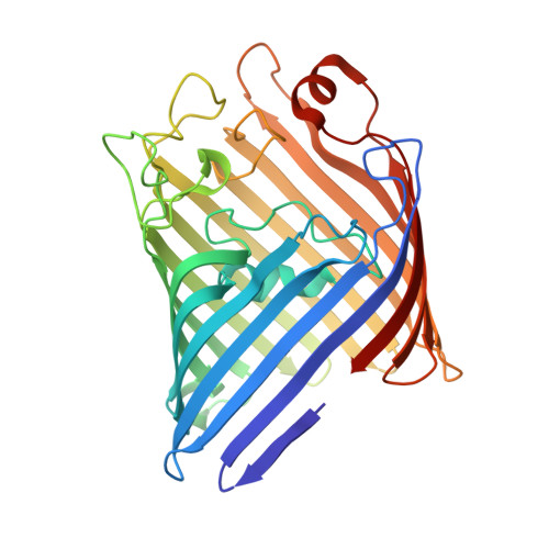
    Porins form transmembrane pores in the outer membrane of Gram-negative bacteria with matrix porin OmpF and osmoporin OmpC from Escherichia coli being differentially expressed depending on environmental conditions. The three-dimensional structure of OmpC has been determined to 2.0 A resolution by X-ray crystallography. As expected from the high sequence similarity, OmpC adopts the OmpF-like 16-stranded hollow beta-barrel fold with three beta-barrels associated to form a tight trimer. Unlike in OmpF, the extracellular loops form a continuous wall at the perimeter of the vestibule common to the three pores, due to a 14-residues insertion in loop L4. The pore constriction and the periplasmic outlet are very similar to OmpF with 74% of the pore lining residues being conserved. Overall, only few ionizable residues are exchanged at the pore lining. The OmpC structure suggests that not pore size, but electrostatic pore potential and particular atomic details of the pore linings are the critical parameters that physiologically distinguish OmpC from OmpF.


  • Organizational Affiliation

    Division of Structural Biology, University of Basel, Klingelbergstrasse 70, CH-4056 Basel, Switzerland.


Macromolecules
Find similar proteins by:  (by identity cutoff)  |  3D Structure
Entity ID: 1
MoleculeChains Sequence LengthOrganismDetailsImage
OUTER MEMBRANE PROTEIN C
A, B, C
346Escherichia coliMutation(s): 0 
Membrane Entity: Yes 
UniProt
Find proteins for P06996 (Escherichia coli (strain K12))
Explore P06996 
Go to UniProtKB:  P06996
Entity Groups  
Sequence Clusters30% Identity50% Identity70% Identity90% Identity95% Identity100% Identity
UniProt GroupP06996
Sequence Annotations
Expand
  • Reference Sequence
Small Molecules
Ligands 3 Unique
IDChains Name / Formula / InChI Key2D Diagram3D Interactions
D12
Query on D12

Download Ideal Coordinates CCD File 
I [auth C]
J [auth C]
K [auth C]
L [auth C]
M [auth C]
I [auth C],
J [auth C],
K [auth C],
L [auth C],
M [auth C],
N [auth C],
O [auth C],
P [auth C],
Q [auth C],
R [auth C],
S [auth C]
DODECANE
C12 H26
SNRUBQQJIBEYMU-UHFFFAOYSA-N
CL
Query on CL

Download Ideal Coordinates CCD File 
F [auth B],
G [auth B]
CHLORIDE ION
Cl
VEXZGXHMUGYJMC-UHFFFAOYSA-M
MG
Query on MG

Download Ideal Coordinates CCD File 
D [auth A],
E [auth B],
H [auth C]
MAGNESIUM ION
Mg
JLVVSXFLKOJNIY-UHFFFAOYSA-N
Experimental Data & Validation

Experimental Data

  • Method: X-RAY DIFFRACTION
  • Resolution: 2.00 Å
  • R-Value Free: 0.214 
  • R-Value Work: 0.180 
  • R-Value Observed: 0.180 
  • Space Group: P 21 21 2
Unit Cell:
Length ( Å )Angle ( ˚ )
a = 200.779α = 90
b = 99.391β = 90
c = 74.328γ = 90
Software Package:
Software NamePurpose
REFMACrefinement
MOSFLMdata reduction
SCALAdata scaling
MOLREPphasing

Structure Validation

View Full Validation Report


Ligand Structure Quality Assessment 


Entry History 

Deposition Data

Revision History  (Full details and data files)

  • Version 1.0: 2006-09-06
    Type: Initial release
  • Version 1.1: 2011-07-13
    Changes: Refinement description, Version format compliance
  • Version 1.2: 2023-12-13
    Changes: Data collection, Database references, Derived calculations, Other, Refinement description