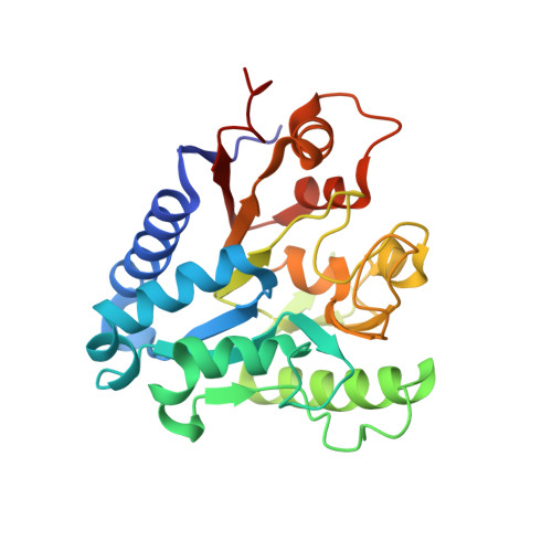
Three Dimensional Structure and Implications for the Catalytic Mechanism of 6-Phosphogluconolactonase from Trypanosoma Brucei.
Delarue, M., Duclert-Savatier, N., Miclet, E., Haouz, A., Giganti, D., Ouazzani, J., Lopez, P., Nilges, M., Stoven, V.(2007) J Mol Biol 366: 868
- PubMed: 17196981
- DOI: https://doi.org/10.1016/j.jmb.2006.11.063
- Primary Citation of Related Structures:
2J0E - PubMed Abstract:
Enzymes from the pentose phosphate pathway (PPP) are potential drug targets for the development of new drugs against Trypanosoma brucei, the causative agent of African sleeping disease: for instance, the 6-phosphogluconate dehydrogenase is currently studied actively for such purposes. Structural and functional studies are necessary to better characterize the associated enzymes and compare them to their human homologues, in order to undertake structure-based drug design studies on such targets. In this context, the crystal structure of 6-phosphogluconolactonase (6PGL) from T. brucei, the second enzyme from PPP, was determined at 2.1 Angstroms resolution. Comparison of its sequence and structure to other related proteins in the 6PGL family with a known structure (Thermotoga maritima Tm6GPL 1PBT and Vibrio cholerae Vc6PGL (1Y89), which have not been discussed in print), or in the glucosamine-6-phosphate-deaminase family (hexameric Escherichia coli 1DEA and monomeric Bacillus subtilis 2BKV), allowed the identification of the 6PGL active site. In addition to the analysis of the crystal structure, 3D NMR interaction studies and docking experiments are reported here. Key residues involved in substrate binding or in catalysis were identified.
Organizational Affiliation:
Unité de Dynamique Structurale des Macromolécules, CNRS URA 2185, Institut Pasteur, 25 Rue du Docteur Roux, 75015 Paris, France. marc.delarue@pasteur.fr