2IVF

Ethylbenzene dehydrogenase from Aromatoleum aromaticum


Experimental Data Snapshot

  • Method: X-RAY DIFFRACTION
  • Resolution: 1.88 Å
  • R-Value Free: 0.183 
  • R-Value Work: 0.148 
  • R-Value Observed: 0.150 

wwPDB Validation   3D Report Full Report


Ligand Structure Quality Assessment 


This is version 1.1 of the entry. See complete history


Literature

Crystal Structure of Ethylbenzene Dehydrogenase from Aromatoleum Aromaticum

Kloer, D.P.Hagel, C.Heider, J.Schulz, G.E.

(2006) Structure 14: 1377

  • DOI: https://doi.org/10.1016/j.str.2006.07.001
  • Primary Citation of Related Structures:  
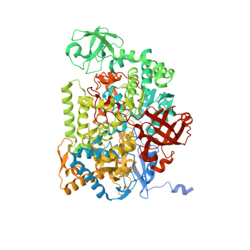
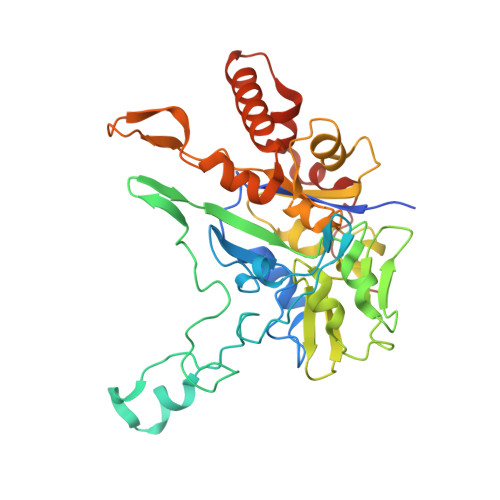
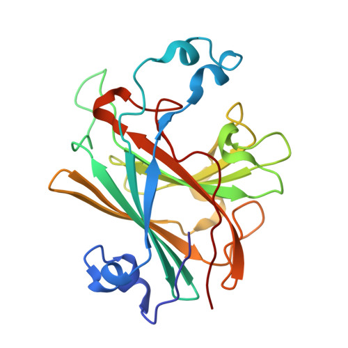
    2IVF

  • PubMed Abstract: 

    Anaerobic degradation of hydrocarbons was discovered a decade ago, and ethylbenzene dehydrogenase was one of the first characterized enzymes involved. The structure of the soluble periplasmic 165 kDa enzyme was established at 1.88 A resolution. It is a heterotrimer. The alpha subunit contains the catalytic center with a molybdenum held by two molybdopterin-guanine dinucleotides, one with an open pyran ring, and an iron-sulfur cluster with a histidine ligand. During catalysis, electrons produced by substrate oxidation are transferred to a heme in the gamma subunit and then presumably to a separate cytochrome involved in nitrate respiration. The beta subunit contains four iron-sulfur clusters and is structurally related to ferredoxins. The gamma subunit is the first known protein with a methionine and a lysine as axial heme ligands. The catalytic product was modeled into the active center, showing the reaction geometry. A mechanism consistent with activity and inhibition data of ethylbenzene-related compounds is proposed.


  • Organizational Affiliation

    Institut für Organische Chemie und Biochemie, Albert-Ludwigs-Universität, Albertstr. 21, D-79104 Freiburg im Breisgau, Germany.


Macromolecules
Find similar proteins by:  (by identity cutoff)  |  3D Structure
Entity ID: 1
MoleculeChains Sequence LengthOrganismDetailsImage
ETHYLBENZENE DEHYDROGENASE ALPHA-SUBUNIT976Aromatoleum aromaticum EbN1Mutation(s): 0 
EC: 1.17.99.2
UniProt
Find proteins for Q5P5I0 (Aromatoleum aromaticum (strain EbN1))
Explore Q5P5I0 
Go to UniProtKB:  Q5P5I0
Entity Groups  
Sequence Clusters30% Identity50% Identity70% Identity90% Identity95% Identity100% Identity
UniProt GroupQ5P5I0
Sequence Annotations
Expand
  • Reference Sequence
Find similar proteins by:  (by identity cutoff)  |  3D Structure
Entity ID: 2
MoleculeChains Sequence LengthOrganismDetailsImage
ETHYLBENZENE DEHYDROGENASE BETA-SUBUNIT352Aromatoleum aromaticum EbN1Mutation(s): 0 
EC: 1.17.99.2
UniProt
Find proteins for Q5P5I1 (Aromatoleum aromaticum (strain EbN1))
Explore Q5P5I1 
Go to UniProtKB:  Q5P5I1
Entity Groups  
Sequence Clusters30% Identity50% Identity70% Identity90% Identity95% Identity100% Identity
UniProt GroupQ5P5I1
Sequence Annotations
Expand
  • Reference Sequence
Find similar proteins by:  (by identity cutoff)  |  3D Structure
Entity ID: 3
MoleculeChains Sequence LengthOrganismDetailsImage
ETHYLBENZENE DEHYDROGENASE GAMMA-SUBUNIT214Aromatoleum aromaticum EbN1Mutation(s): 0 
EC: 1.17.99.2
UniProt
Find proteins for Q5P5I2 (Aromatoleum aromaticum (strain EbN1))
Explore Q5P5I2 
Go to UniProtKB:  Q5P5I2
Entity Groups  
Sequence Clusters30% Identity50% Identity70% Identity90% Identity95% Identity100% Identity
UniProt GroupQ5P5I2
Sequence Annotations
Expand
  • Reference Sequence
Small Molecules
Ligands 10 Unique
IDChains Name / Formula / InChI Key2D Diagram3D Interactions
MD1
Query on MD1

Download Ideal Coordinates CCD File 
N [auth A]PHOSPHORIC ACID 4-(2-AMINO-4-OXO-3,4,5,6,-TETRAHYDRO-PTERIDIN-6-YL)-2-HYDROXY-3,4-DIMERCAPTO-BUT-3-EN-YL ESTER GUANYLATE ESTER
C20 H26 N10 O13 P2 S2
IRGDLSAXQOKWLX-XHEYTWMPSA-N
MGD
Query on MGD

Download Ideal Coordinates CCD File 
M [auth A]2-AMINO-5,6-DIMERCAPTO-7-METHYL-3,7,8A,9-TETRAHYDRO-8-OXA-1,3,9,10-TETRAAZA-ANTHRACEN-4-ONE GUANOSINE DINUCLEOTIDE
C20 H26 N10 O13 P2 S2
VQAGYJCYOLHZDH-ILXWUORBSA-N
HEM
Query on HEM

Download Ideal Coordinates CCD File 
X [auth C]PROTOPORPHYRIN IX CONTAINING FE
C34 H32 Fe N4 O4
KABFMIBPWCXCRK-RGGAHWMASA-L
SF4
Query on SF4

Download Ideal Coordinates CCD File 
K [auth A],
S [auth B],
T [auth B],
U [auth B]
IRON/SULFUR CLUSTER
Fe4 S4
LJBDFODJNLIPKO-UHFFFAOYSA-N
F3S
Query on F3S

Download Ideal Coordinates CCD File 
R [auth B]FE3-S4 CLUSTER
Fe3 S4
FCXHZBQOKRZXKS-UHFFFAOYSA-N
MES
Query on MES

Download Ideal Coordinates CCD File 
D [auth A]2-(N-MORPHOLINO)-ETHANESULFONIC ACID
C6 H13 N O4 S
SXGZJKUKBWWHRA-UHFFFAOYSA-N
MO
Query on MO

Download Ideal Coordinates CCD File 
L [auth A]MOLYBDENUM ATOM
Mo
ZOKXTWBITQBERF-UHFFFAOYSA-N
PO4
Query on PO4

Download Ideal Coordinates CCD File 
P [auth B]PHOSPHATE ION
O4 P
NBIIXXVUZAFLBC-UHFFFAOYSA-K
GOL
Query on GOL

Download Ideal Coordinates CCD File 
F [auth A]
G [auth A]
H [auth A]
I [auth A]
O [auth B]
F [auth A],
G [auth A],
H [auth A],
I [auth A],
O [auth B],
Q [auth B],
V [auth C],
W [auth C]
GLYCEROL
C3 H8 O3
PEDCQBHIVMGVHV-UHFFFAOYSA-N
ACT
Query on ACT

Download Ideal Coordinates CCD File 
E [auth A],
J [auth A]
ACETATE ION
C2 H3 O2
QTBSBXVTEAMEQO-UHFFFAOYSA-M
Experimental Data & Validation

Experimental Data

  • Method: X-RAY DIFFRACTION
  • Resolution: 1.88 Å
  • R-Value Free: 0.183 
  • R-Value Work: 0.148 
  • R-Value Observed: 0.150 
  • Space Group: P 1 21 1
Unit Cell:
Length ( Å )Angle ( ˚ )
a = 112.532α = 90
b = 67.324β = 111.3
c = 114.765γ = 90
Software Package:
Software NamePurpose
REFMACrefinement
XDSdata reduction
XSCALEdata scaling
MLPHAREphasing

Structure Validation

View Full Validation Report


Ligand Structure Quality Assessment 


Entry History 

Deposition Data

Revision History  (Full details and data files)

  • Version 1.0: 2006-09-14
    Type: Initial release
  • Version 1.1: 2011-07-13
    Changes: Advisory, Source and taxonomy, Version format compliance