2IS3

Crystal Structure of Escherichia coli RNase T


Experimental Data Snapshot

  • Method: X-RAY DIFFRACTION
  • Resolution: 3.10 Å
  • R-Value Free: 0.231 
  • R-Value Work: 0.196 
  • R-Value Observed: 0.197 

wwPDB Validation   3D Report Full Report


This is version 1.3 of the entry. See complete history


Literature

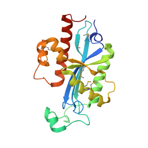
Crystal Structure of RNase T, an Exoribonuclease Involved in tRNA Maturation and End Turnover.

Zuo, Y.Zheng, H.Wang, Y.Chruszcz, M.Cymborowski, M.Skarina, T.Savchenko, A.Malhotra, A.Minor, W.

(2007) Structure 15: 417-428

  • DOI: https://doi.org/10.1016/j.str.2007.02.004
  • Primary Citation of Related Structures:  
    2F96, 2IS3

  • PubMed Abstract: 

    The 3' processing of most bacterial precursor tRNAs involves exonucleolytic trimming to yield a mature CCA end. This step is carried out by RNase T, a member of the large DEDD family of exonucleases. We report the crystal structures of RNase T from Escherichia coli and Pseudomonas aeruginosa, which show that this enzyme adopts an opposing dimeric arrangement, with the catalytic DEDD residues from one monomer closely juxtaposed with a large basic patch on the other monomer. This arrangement suggests that RNase T has to be dimeric for substrate specificity, and agrees very well with prior site-directed mutagenesis studies. The dimeric architecture of RNase T is very similar to the arrangement seen in oligoribonuclease, another bacterial DEDD family exoribonuclease. The catalytic residues in these two enzymes are organized very similarly to the catalytic domain of the third DEDD family exoribonuclease in E. coli, RNase D, which is monomeric.


  • Organizational Affiliation

    Department of Biochemistry and Molecular Biology, University of Miami School of Medicine, Miami, FL 33101, USA.


Macromolecules
Find similar proteins by:  (by identity cutoff)  |  3D Structure
Entity ID: 1
MoleculeChains Sequence LengthOrganismDetailsImage
Ribonuclease T
A, B, C, D
215Escherichia coli K-12Mutation(s): 7 
Gene Names: rnt
EC: 3.1.13
UniProt
Find proteins for P30014 (Escherichia coli (strain K12))
Explore P30014 
Go to UniProtKB:  P30014
Entity Groups  
Sequence Clusters30% Identity50% Identity70% Identity90% Identity95% Identity100% Identity
UniProt GroupP30014
Sequence Annotations
Expand
  • Reference Sequence
Small Molecules
Ligands 1 Unique
IDChains Name / Formula / InChI Key2D Diagram3D Interactions
SO4
Query on SO4

Download Ideal Coordinates CCD File 
E [auth A]
F [auth A]
G [auth A]
H [auth B]
I [auth B]
E [auth A],
F [auth A],
G [auth A],
H [auth B],
I [auth B],
J [auth B],
K [auth B],
L [auth C],
M [auth C],
N [auth C],
O [auth D],
P [auth D],
Q [auth D]
SULFATE ION
O4 S
QAOWNCQODCNURD-UHFFFAOYSA-L
Modified Residues  1 Unique
IDChains TypeFormula2D DiagramParent
MSE
Query on MSE
A, B, C, D
L-PEPTIDE LINKINGC5 H11 N O2 SeMET
Experimental Data & Validation

Experimental Data

  • Method: X-RAY DIFFRACTION
  • Resolution: 3.10 Å
  • R-Value Free: 0.231 
  • R-Value Work: 0.196 
  • R-Value Observed: 0.197 
  • Space Group: I 4 2 2
Unit Cell:
Length ( Å )Angle ( ˚ )
a = 213.14α = 90
b = 213.14β = 90
c = 149.15γ = 90
Software Package:
Software NamePurpose
REFMACrefinement
CBASSdata collection
DENZOdata reduction
SCALEPACKdata scaling
SnBphasing

Structure Validation

View Full Validation Report


Entry History 

Deposition Data

Revision History  (Full details and data files)

  • Version 1.0: 2007-04-24
    Type: Initial release
  • Version 1.1: 2008-05-01
    Changes: Version format compliance
  • Version 1.2: 2011-07-13
    Changes: Advisory, Version format compliance
  • Version 1.3: 2017-10-18
    Changes: Refinement description