2IPI

Crystal Structure of Aclacinomycin Oxidoreductase


Experimental Data Snapshot

  • Method: X-RAY DIFFRACTION
  • Resolution: 1.65 Å
  • R-Value Free: 0.242 
  • R-Value Observed: 0.185 

wwPDB Validation   3D Report Full Report


Ligand Structure Quality Assessment 


This is version 1.2 of the entry. See complete history


Literature

Structure determination by multiwavelength anomalous diffraction of aclacinomycin oxidoreductase: indications of multidomain pseudomerohedral twinning.

Sultana, A.Alexeev, I.Kursula, I.Mantsala, P.Niemi, J.Schneider, G.

(2007) Acta Crystallogr D Biol Crystallogr 63: 149-159

  • DOI: https://doi.org/10.1107/S0907444906044271
  • Primary Citation of Related Structures:  
    2IPI

  • PubMed Abstract: 

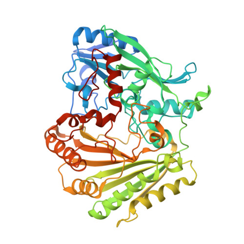
    The crystal structure of aclacinomycin oxidoreductase (AknOx), a tailoring enzyme involved in the biosynthesis of the polyketide antibiotic aclacinomycin, was determined to 1.65 A resolution by multiwavelength anomalous diffraction using data from selenomethionine-substituted crystals. The crystals belong to space group P2(1), with unit-cell parameters a = 68.2, b = 264.5, c = 68.2 A, beta = 119 degrees . Analysis of the intensity statistics clearly showed the presence of pseudomerohedral twinning. The data set could also be indexed and scaled with an R(sym) of 0.072 in the orthorhombic space group C222(1) (unit-cell parameters a = 69.7, b = 117.5, c = 264.4 A), indicating the possibility of pseudomerohedral twinning along the diagonal between the monoclinic a and c directions. Refinement using this twin operator resulted in an R(free) of 24.2%. A monoclinic lattice with a = c and beta close to 120 degrees can emulate a hexagonal metric, with the possibility of a threefold twin operator along the b axis and three twin domains. Refinement assuming three-domain twinning gave a final R(free) of 26.5%. The structure of AknOx can be thus refined with comparable R(free) values using either of the twin operators separately, suggesting the possibility that crystals of AknOx contain six twin domains generated by the twofold and threefold twin operators perpendicular to each other. Both twin operators coincide with noncrystallographic symmetry axes that may promote twinning.


  • Organizational Affiliation

    Department of Medical Biochemistry and Biophysics, Karolinska Institutet, S-171 77 Stockholm, Sweden.


Macromolecules
Find similar proteins by:  (by identity cutoff)  |  3D Structure
Entity ID: 1
MoleculeChains Sequence LengthOrganismDetailsImage
Aclacinomycin oxidoreductase (AknOx)
A, B, C, D
521Streptomyces galilaeusMutation(s): 0 
Gene Names: AknOx
UniProt
Find proteins for Q0PCD7 (Streptomyces galilaeus)
Explore Q0PCD7 
Go to UniProtKB:  Q0PCD7
Entity Groups  
Sequence Clusters30% Identity50% Identity70% Identity90% Identity95% Identity100% Identity
UniProt GroupQ0PCD7
Sequence Annotations
Expand
  • Reference Sequence
Small Molecules
Ligands 2 Unique
IDChains Name / Formula / InChI Key2D Diagram3D Interactions
AKY
Query on AKY

Download Ideal Coordinates CCD File 
E [auth A]METHYL (2S,4R)-2-ETHYL-2,5,7-TRIHYDROXY-6,11-DIOXO-4-{[2,3,6-TRIDEOXY-4-O-{2,6-DIDEOXY-4-O-[(2S,6S)-6-METHYL-5-OXOTETRAHYDRO-2H -PYRAN-2-YL]-ALPHA-D-LYXO-HEXOPYRANOSYL}-3-(DIMETHYLAMINO)-D-RIBO-HEXOPYRANOSYL]OXY}-1,2,3,4,6,11-HEXAHYDROTETRACENE-1-C ARBOXYLATE
C42 H53 N O15
USZYSDMBJDPRIF-JAFKFOJZSA-N
FAD
Query on FAD

Download Ideal Coordinates CCD File 
F [auth A],
G [auth B],
H [auth C],
I [auth D]
FLAVIN-ADENINE DINUCLEOTIDE
C27 H33 N9 O15 P2
VWWQXMAJTJZDQX-UYBVJOGSSA-N
Experimental Data & Validation

Experimental Data

  • Method: X-RAY DIFFRACTION
  • Resolution: 1.65 Å
  • R-Value Free: 0.242 
  • R-Value Observed: 0.185 
  • Space Group: P 1 21 1
Unit Cell:
Length ( Å )Angle ( ˚ )
a = 68.5α = 90
b = 266.2β = 119
c = 68.7γ = 90
Software Package:
Software NamePurpose
SOLVEphasing
RESOLVEphasing
REFMACrefinement
PDB_EXTRACTdata extraction
XDSdata reduction
XSCALEdata scaling
SHELXL-97refinement

Structure Validation

View Full Validation Report


Ligand Structure Quality Assessment 


Entry History 

Deposition Data

Revision History  (Full details and data files)

  • Version 1.0: 2007-01-23
    Type: Initial release
  • Version 1.1: 2008-05-01
    Changes: Version format compliance
  • Version 1.2: 2011-07-13
    Changes: Version format compliance