2INP

Structure of the Phenol Hydroxylase-Regulatory Protein Complex


Experimental Data Snapshot

  • Method: X-RAY DIFFRACTION
  • Resolution: 2.30 Å
  • R-Value Free: 0.236 
  • R-Value Work: 0.192 
  • R-Value Observed: 0.198 

wwPDB Validation   3D Report Full Report


This is version 1.3 of the entry. See complete history


Literature

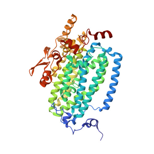
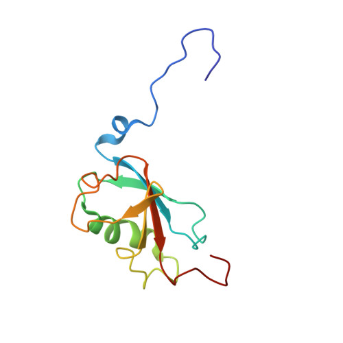
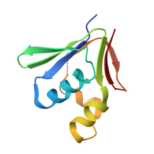
X-ray Structure of a Hydroxylase-Regulatory Protein Complex from a Hydrocarbon-Oxidizing Multicomponent Monooxygenase, Pseudomonas sp. OX1 Phenol Hydroxylase.

Sazinsky, M.H.Dunten, P.W.McCormick, M.S.Didonato, A.Lippard, S.J.

(2006) Biochemistry 45: 15392-15404

  • DOI: https://doi.org/10.1021/bi0618969
  • Primary Citation of Related Structures:  
    2INN, 2INP

  • PubMed Abstract: 

    Phenol hydroxylase (PH) belongs to a family of bacterial multicomponent monooxygenases (BMMs) with carboxylate-bridged diiron active sites. Included are toluene/o-xylene (ToMO) and soluble methane (sMMO) monooxygenase. PH hydroxylates aromatic compounds, but unlike sMMO, it cannot oxidize alkanes despite having a similar dinuclear iron active site. Important for activity is formation of a complex between the hydroxylase and a regulatory protein component. To address how structural features of BMM hydroxylases and their component complexes may facilitate the catalytic mechanism and choice of substrate, we determined X-ray structures of native and SeMet forms of the PH hydroxylase (PHH) in complex with its regulatory protein (PHM) to 2.3 A resolution. PHM binds in a canyon on one side of the (alphabetagamma)2 PHH dimer, contacting alpha-subunit helices A, E, and F approximately 12 A above the diiron core. The structure of the dinuclear iron center in PHH resembles that of mixed-valent MMOH, suggesting an Fe(II)Fe(III) oxidation state. Helix E, which comprises part of the iron-coordinating four-helix bundle, has more pi-helical character than analogous E helices in MMOH and ToMOH lacking a bound regulatory protein. Consequently, conserved active site Thr and Asn residues translocate to the protein surface, and an approximately 6 A pore opens through the four-helix bundle. Of likely functional significance is a specific hydrogen bond formed between this Asn residue and a conserved Ser side chain on PHM. The PHM protein covers a putative docking site on PHH for the PH reductase, which transfers electrons to the PHH diiron center prior to O2 activation, suggesting that the regulatory component may function to block undesired reduction of oxygenated intermediates during the catalytic cycle. A series of hydrophobic cavities through the PHH alpha-subunit, analogous to those in MMOH, may facilitate movement of the substrate to and/or product from the active site pocket. Comparisons between the ToMOH and PHH structures provide insights into their substrate regiospecificities.


  • Organizational Affiliation

    Department of Chemistry, Massachusetts Institute of Technology, Cambridge, Massachusetts 02139, USA.


Macromolecules
Find similar proteins by:  (by identity cutoff)  |  3D Structure
Entity ID: 1
MoleculeChains Sequence LengthOrganismDetailsImage
Phenol hydroxylase component phN
A, B
494Stutzerimonas stutzeriMutation(s): 0 
Gene Names: phN
UniProt
Find proteins for Q84AQ2 (Stutzerimonas stutzeri)
Explore Q84AQ2 
Go to UniProtKB:  Q84AQ2
Entity Groups  
Sequence Clusters30% Identity50% Identity70% Identity90% Identity95% Identity100% Identity
UniProt GroupQ84AQ2
Sequence Annotations
Expand
  • Reference Sequence
Find similar proteins by:  (by identity cutoff)  |  3D Structure
Entity ID: 2
MoleculeChains Sequence LengthOrganismDetailsImage
Phenol hydroxylase component phL
C, D
328Stutzerimonas stutzeriMutation(s): 0 
Gene Names: phL
UniProt
Find proteins for Q84AQ4 (Stutzerimonas stutzeri)
Explore Q84AQ4 
Go to UniProtKB:  Q84AQ4
Entity Groups  
Sequence Clusters30% Identity50% Identity70% Identity90% Identity95% Identity100% Identity
UniProt GroupQ84AQ4
Sequence Annotations
Expand
  • Reference Sequence
Find similar proteins by:  (by identity cutoff)  |  3D Structure
Entity ID: 3
MoleculeChains Sequence LengthOrganismDetailsImage
Phenol hydroxylase component phO
E, F
118Stutzerimonas stutzeriMutation(s): 0 
Gene Names: phO
UniProt
Find proteins for Q84AQ1 (Stutzerimonas stutzeri)
Explore Q84AQ1 
Go to UniProtKB:  Q84AQ1
Entity Groups  
Sequence Clusters30% Identity50% Identity70% Identity90% Identity95% Identity100% Identity
UniProt GroupQ84AQ1
Sequence Annotations
Expand
  • Reference Sequence
Find similar proteins by:  (by identity cutoff)  |  3D Structure
Entity ID: 4
MoleculeChains Sequence LengthOrganismDetailsImage
Phenol hydroxylase component phMG [auth L]89Stutzerimonas stutzeriMutation(s): 0 
Gene Names: phM
UniProt
Find proteins for Q84AQ3 (Stutzerimonas stutzeri)
Explore Q84AQ3 
Go to UniProtKB:  Q84AQ3
Entity Groups  
Sequence Clusters30% Identity50% Identity70% Identity90% Identity95% Identity100% Identity
UniProt GroupQ84AQ3
Sequence Annotations
Expand
  • Reference Sequence
Experimental Data & Validation

Experimental Data

  • Method: X-RAY DIFFRACTION
  • Resolution: 2.30 Å
  • R-Value Free: 0.236 
  • R-Value Work: 0.192 
  • R-Value Observed: 0.198 
  • Space Group: P 21 21 21
Unit Cell:
Length ( Å )Angle ( ˚ )
a = 87.751α = 90
b = 146.305β = 90
c = 190.02γ = 90
Software Package:
Software NamePurpose
REFMACrefinement
PDB_EXTRACTdata extraction
ADSCdata collection
HKL-2000data reduction
HKL-2000data scaling
CNSphasing

Structure Validation

View Full Validation Report


Entry History 

Deposition Data

Revision History  (Full details and data files)

  • Version 1.0: 2007-01-16
    Type: Initial release
  • Version 1.1: 2008-05-01
    Changes: Version format compliance
  • Version 1.2: 2011-07-13
    Changes: Version format compliance
  • Version 1.3: 2023-08-30
    Changes: Data collection, Database references, Derived calculations, Refinement description