Crystal structure of uroporphyrinogen decarboxylase from Bacillus subtilis
Fan, J., Liu, Q., Hao, Q., Teng, M.K., Niu, L.W.(2007) J Bacteriol 189: 3573-3580
- PubMed: 17122346
- DOI: https://doi.org/10.1128/JB.01083-06
- Primary Citation of Related Structures:
2INF - PubMed Abstract:
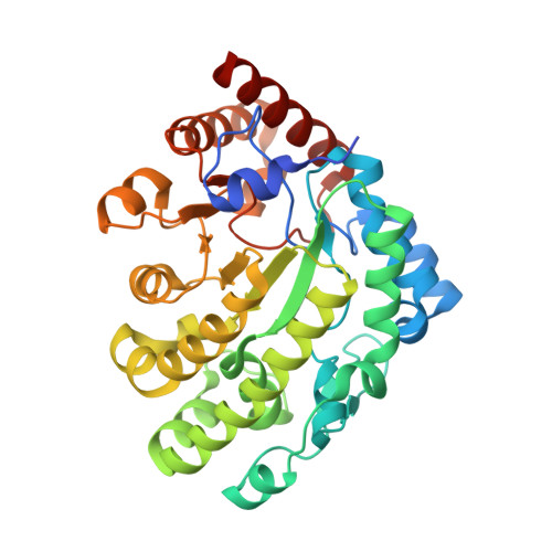
Uroporphyrinogen decarboxylase (UROD) is a branch point enzyme in the biosynthesis of the tetrapyrroles. It catalyzes the decarboxylation of four acetate groups of uroporphyrinogen III to yield coproporphyrinogen III, leading to heme and chlorophyll biosynthesis. UROD is a special type of nonoxidative decarboxylase, since no cofactor is essential for catalysis. In this work, the first crystal structure of a bacterial UROD, Bacillus subtilis UROD (UROD(Bs)), has been determined at a 2.3 A resolution. The biological unit of UROD(Bs) was determined by dynamic light scattering measurements to be a homodimer in solution. There are four molecules in the crystallographic asymmetric unit, corresponding to two homodimers. Structural comparison of UROD(Bs) with eukaryotic URODs reveals a variation of two loops, which possibly affect the binding of substrates and release of products. Structural comparison with the human UROD-coproporphyrinogen III complex discloses a similar active cleft, with five invariant polar residues (Arg29, Arg33, Asp78, Tyr154, and His322) and three invariant hydrophobic residues (Ile79, Phe144, and Phe207), in UROD(Bs). Among them, Asp78 may interact with the pyrrole NH groups of the substrate, and Arg29 is a candidate for positioning the acetate groups of the substrate. Both residues may also play catalytic roles.
Organizational Affiliation:
Hefei National laboratory of Physical Sciences at Microscale and School of Life Sciences, University of Science & Technology of China, Hefei Anhui, 230027, China.