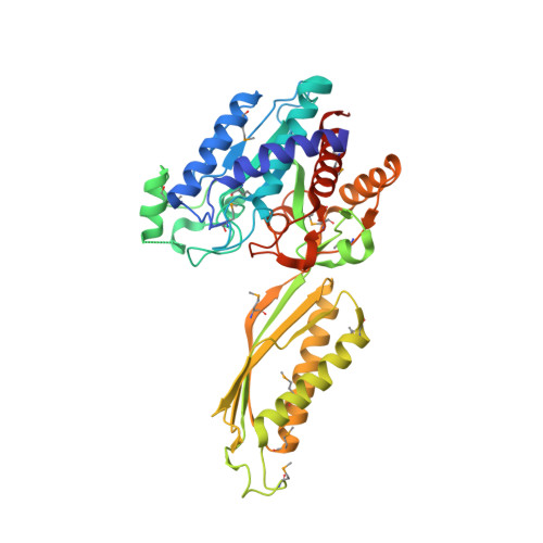
Structural Analysis of a Ternary Complex of Allantoate Amidohydrolase from Escherichia coli Reveals its Mechanics.
Agarwal, R., Burley, S.K., Swaminathan, S.(2007) J Mol Biol 368: 450-463
- PubMed: 17362992
- DOI: https://doi.org/10.1016/j.jmb.2007.02.028
- Primary Citation of Related Structures:
1Z2L, 2IMO - PubMed Abstract:
Purine metabolism plays a major role in regulating the availability of purine nucleotides destined for nucleic acid synthesis. Allantoate amidohydrolase catalyzes the conversion of allantoate to (S)-ureidoglycolate, one of the crucial alternate steps in purine metabolism. The crystal structure of a ternary complex of allantoate amidohydrolase with its substrate allantoate and an allosteric effector, a sulfate ion, from Escherichia coli was determined to understand better the catalytic mechanism and substrate specificity. The 2.25 A resolution X-ray structure reveals an alpha/beta scaffold akin to zinc exopeptidases of the peptidase M20 family and lacks the (beta/alpha)(8)-barrel fold characteristic of the amidohydrolases. Arrangement of the substrate and the two co-catalytic zinc ions at the active site governs catalytic specificity for hydrolysis of N-carbamyl versus the peptide bond in exopeptidases. In its crystalline form, allantoate amidohydrolase adopts a relatively open conformation. However, structural analysis reveals the possibility of a significant movement of domains via rotation about two hinge regions upon allosteric effector and substrate binding resulting in a closed catalytically competent conformation by bringing the substrate allantoate closer to co-catalytic zinc ions. Two cis-prolyl peptide bonds found on either side of the dimerization domain in close proximity to the substrate and ligand-binding sites may be involved in protein folding and in preserving the integrity of the catalytic site.
Organizational Affiliation:
Biology Department, Brookhaven National Laboratory, Upton, NY 11973, USA.