2ILN

Crystal structure of the Bowman-Birk inhibitor from snail medic seeds in complex with bovine trypsin


Experimental Data Snapshot

  • Method: X-RAY DIFFRACTION
  • Resolution: 2.00 Å
  • R-Value Free: 0.287 
  • R-Value Work: 0.232 
  • R-Value Observed: 0.235 

wwPDB Validation   3D Report Full Report


This is version 1.4 of the entry. See complete history


Literature

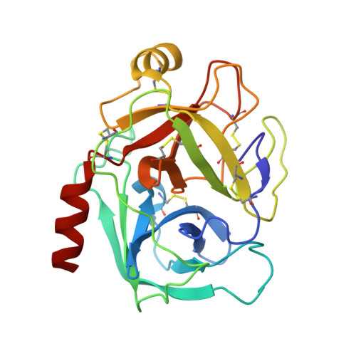
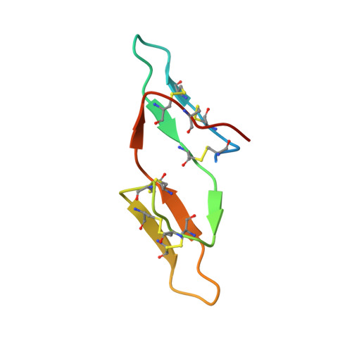
Crystal structure of the anticarcinogenic Bowman-Birk inhibitor from snail medic (Medicago scutellata) seeds complexed with bovine trypsin

Capaldi, S.Perduca, M.Faggion, B.Carrizo, M.E.Tava, A.Ragona, L.Monaco, H.L.

(2007) J Struct Biol 158: 71-79

  • DOI: https://doi.org/10.1016/j.jsb.2006.10.017
  • Primary Citation of Related Structures:  
    2ILN

  • PubMed Abstract: 

    The structure of the ternary complex of the anticarcinogenic Bowman-Birk protease inhibitor purified from snail medic (Medicago scutellata) seeds (MSTI) and two molecules of bovine trypsin has been solved by X-ray diffraction analysis of single crystals to a resolution of 2.0 A. This is the highest resolution model of a ternary complex of this type currently available. The two binding loops of the MSTI differ in only one amino acid and have in both cases an arginine in position P1. The distances between the residues of the inhibitor at the binding interface and the trypsin side chains that recognize them are almost identical in the two sites. When compared to the NMR model of the uncomplexed MSTI, the inhibitor in the functional assembly with trypsin shows the largest differences in the two P2' residues. Compared with the similar ternary complex of the soybean trypsin inhibitor, this model shows very small differences in the polypeptide chain of the trypsin binding sites and its largest difference in the area between Asp 26 and His 32 of the MSTI which in the soybean inhibitor has an extra Leu inserted in position 29.


  • Organizational Affiliation

    Biocrystallography Laboratory, Department of Science and Technology, University of Verona, Strada Le Grazie 15, 37134 Verona, Italy.


Macromolecules
Find similar proteins by:  (by identity cutoff)  |  3D Structure
Entity ID: 1
MoleculeChains Sequence LengthOrganismDetailsImage
Cationic trypsin
A, B
223Bos taurusMutation(s): 0 
EC: 3.4.21.4
UniProt
Find proteins for P00760 (Bos taurus)
Explore P00760 
Go to UniProtKB:  P00760
Entity Groups  
Sequence Clusters30% Identity50% Identity70% Identity90% Identity95% Identity100% Identity
UniProt GroupP00760
Sequence Annotations
Expand
  • Reference Sequence
Find similar proteins by:  (by identity cutoff)  |  3D Structure
Entity ID: 2
MoleculeChains Sequence LengthOrganismDetailsImage
Bowman-Birk type proteinase inhibitorC [auth I]62Medicago scutellataMutation(s): 0 
UniProt
Find proteins for P80321 (Medicago scutellata)
Explore P80321 
Go to UniProtKB:  P80321
Entity Groups  
Sequence Clusters30% Identity50% Identity70% Identity90% Identity95% Identity100% Identity
UniProt GroupP80321
Sequence Annotations
Expand
  • Reference Sequence
Experimental Data & Validation

Experimental Data

  • Method: X-RAY DIFFRACTION
  • Resolution: 2.00 Å
  • R-Value Free: 0.287 
  • R-Value Work: 0.232 
  • R-Value Observed: 0.235 
  • Space Group: P 41
Unit Cell:
Length ( Å )Angle ( ˚ )
a = 54.47α = 90
b = 54.47β = 90
c = 180.08γ = 90
Software Package:
Software NamePurpose
REFMACrefinement
MAR345data collection
MOSFLMdata reduction
CCP4data scaling
MOLREPphasing

Structure Validation

View Full Validation Report


Entry History 

Deposition Data

Revision History  (Full details and data files)

  • Version 1.0: 2007-04-10
    Type: Initial release
  • Version 1.1: 2008-05-01
    Changes: Version format compliance
  • Version 1.2: 2011-07-13
    Changes: Version format compliance
  • Version 1.3: 2017-10-18
    Changes: Refinement description
  • Version 1.4: 2023-10-25
    Changes: Data collection, Database references, Refinement description