2IID

Structure of L-amino acid oxidase from Calloselasma rhodostoma in complex with L-phenylalanine


Experimental Data Snapshot

  • Method: X-RAY DIFFRACTION
  • Resolution: 1.80 Å
  • R-Value Free: 0.210 
  • R-Value Work: 0.172 
  • R-Value Observed: 0.174 

wwPDB Validation   3D Report Full Report


This is version 2.1 of the entry. See complete history


Literature

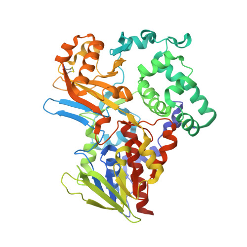
Crystal Structure of LAAO from Calloselasma rhodostoma with an L-Phenylalanine Substrate: Insights into Structure and Mechanism

Moustafa, I.M.Foster, S.Lyubimov, A.Y.Vrielink, A.

(2006) J Mol Biol 364: 991-1002

  • DOI: https://doi.org/10.1016/j.jmb.2006.09.032
  • Primary Citation of Related Structures:  
    2IID

  • PubMed Abstract: 

    L-Amino acid oxidase is a dimeric glycosylated flavoenzyme, a major constituent of the venom-from the snake Calloselasma rhodostoma. The enzyme exhibits apoptosis inducing effects as well as antibacterial and anti-HIV activities. The structure of l-amino acid oxidase with its substrate (L-phenylalanine) has been refined to a resolution of 1.8 A. The complex structure reveals the substrate bound to the reduced flavin (FADred). Alternative conformations for the key residues His223 and Arg322 are evident, suggesting a dynamic active site. Furthermore, conformational changes are apparent for the isoalloxazine ring; the three-ring system exhibits more bending around the N5-N10 axis compared to the oxidized flavin. The implications of the observed dynamics on the mechanism of catalysis are discussed. Inspection of buried surfaces in the enzyme reveals a Y-shaped channel system extending from the external surface of the protein to the active site. One portion of this channel may serve as the entry path for O2 during the oxidative half-reaction. The second region, separated from the proposed O2 channel by the N terminus (residues 8-16) of the protein, may play a role in H2O2 release. Interestingly, the latter portion of the channel would direct the H2O2 product to the exterior surface of the protein, near the glycan moiety, thought to anchor the enzyme to the host cell. This channel location may explain the ability of the enzyme to localize H2O2 to the targeted cell and thus induce the apoptotic effect.


  • Organizational Affiliation

    Department of Chemistry and Biochemistry, University of California, Santa Cruz, Santa Cruz, CA 95064, USA.


Macromolecules
Find similar proteins by:  (by identity cutoff)  |  3D Structure
Entity ID: 1
MoleculeChains Sequence LengthOrganismDetailsImage
L-amino-acid oxidase
A, B, C, D
498Calloselasma rhodostomaMutation(s): 0 
EC: 1.4.3.2
UniProt
Find proteins for P81382 (Calloselasma rhodostoma)
Explore P81382 
Go to UniProtKB:  P81382
Entity Groups  
Sequence Clusters30% Identity50% Identity70% Identity90% Identity95% Identity100% Identity
UniProt GroupP81382
Sequence Annotations
Expand
  • Reference Sequence
Oligosaccharides

Help

Entity ID: 2
MoleculeChains Length2D Diagram Glycosylation3D Interactions
alpha-L-fucopyranose-(1-6)-2-acetamido-2-deoxy-beta-D-glucopyranose
E, F, G
2N-Glycosylation
Glycosylation Resources
GlyTouCan:  G86851RC
GlyCosmos:  G86851RC
GlyGen:  G86851RC
Small Molecules
Ligands 3 Unique
IDChains Name / Formula / InChI Key2D Diagram3D Interactions
FAD
Query on FAD

Download Ideal Coordinates CCD File 
J [auth A],
N [auth B],
Q [auth C],
T [auth D]
FLAVIN-ADENINE DINUCLEOTIDE
C27 H33 N9 O15 P2
VWWQXMAJTJZDQX-UYBVJOGSSA-N
NAG
Query on NAG

Download Ideal Coordinates CCD File 
H [auth A],
K [auth B],
L [auth B],
O [auth C],
R [auth D]
2-acetamido-2-deoxy-beta-D-glucopyranose
C8 H15 N O6
OVRNDRQMDRJTHS-FMDGEEDCSA-N
PHE
Query on PHE

Download Ideal Coordinates CCD File 
I [auth A],
M [auth B],
P [auth C],
S [auth D]
PHENYLALANINE
C9 H11 N O2
COLNVLDHVKWLRT-QMMMGPOBSA-N
Experimental Data & Validation

Experimental Data

  • Method: X-RAY DIFFRACTION
  • Resolution: 1.80 Å
  • R-Value Free: 0.210 
  • R-Value Work: 0.172 
  • R-Value Observed: 0.174 
  • Space Group: P 1 21 1
Unit Cell:
Length ( Å )Angle ( ˚ )
a = 78.76α = 90
b = 154.003β = 109.52
c = 103.183γ = 90
Software Package:
Software NamePurpose
REFMACrefinement
HKL-2000data reduction
SCALEPACKdata scaling

Structure Validation

View Full Validation Report


Entry History 

Deposition Data

Revision History  (Full details and data files)

  • Version 1.0: 2006-10-31
    Type: Initial release
  • Version 1.1: 2007-10-16
    Changes: Version format compliance
  • Version 1.2: 2011-07-13
    Changes: Non-polymer description, Version format compliance
  • Version 2.0: 2020-07-29
    Type: Remediation
    Reason: Carbohydrate remediation
    Changes: Atomic model, Data collection, Derived calculations, Structure summary
  • Version 2.1: 2023-08-30
    Changes: Data collection, Database references, Refinement description, Structure summary