2IBN

Crystal structure of Human myo-Inositol Oxygenase (MIOX)


Experimental Data Snapshot

  • Method: X-RAY DIFFRACTION
  • Resolution: 1.50 Å
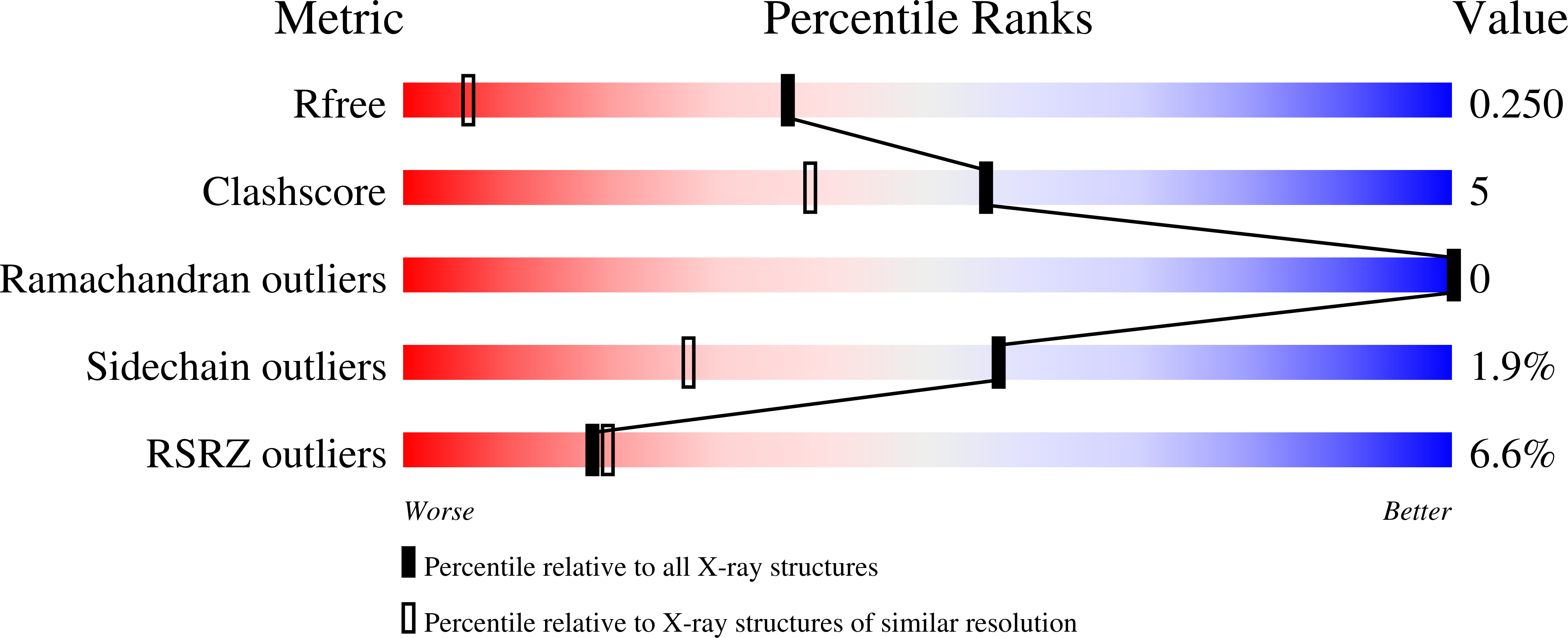
  • R-Value Free: 0.248 
  • R-Value Work: 0.206 
  • R-Value Observed: 0.208 

wwPDB Validation   3D Report Full Report


Ligand Structure Quality Assessment 


This is version 1.4 of the entry. See complete history


Literature

Structural and Biophysical Characterization of Human myo-Inositol Oxygenase

Thorsell, A.G.Persson, C.Voevodskaya, N.Busam, R.D.Hammarstrom, M.Graslund, S.Graslund, A.Hallberg, B.M.

(2008) J Biol Chem 283: 15209-15216

  • DOI: https://doi.org/10.1074/jbc.M800348200
  • Primary Citation of Related Structures:  
    2IBN, 3BXD

  • PubMed Abstract: 

    Altered inositol metabolism is implicated in a number of diabetic complications. The first committed step in mammalian inositol catabolism is performed by myo-inositol oxygenase (MIOX), which catalyzes a unique four-electron dioxygen-dependent ring cleavage of myo-inositol to D-glucuronate. Here, we present the crystal structure of human MIOX in complex with myo-inosose-1 bound in a terminal mode to the MIOX diiron cluster site. Furthermore, from biochemical and biophysical results from N-terminal deletion mutagenesis we show that the N terminus is important, through coordination of a set of loops covering the active site, in shielding the active site during catalysis. EPR spectroscopy of the unliganded enzyme displays a two-component spectrum that we can relate to an open and a closed active site conformation. Furthermore, based on site-directed mutagenesis in combination with biochemical and biophysical data, we propose a novel role for Lys(127) in governing access to the diiron cluster.


  • Organizational Affiliation

    Department of Cell and Molecular Biology, Medical Nobel Institute, Karolinska Institutet, SE-171 77 Stockholm, Sweden.


Macromolecules
Find similar proteins by:  (by identity cutoff)  |  3D Structure
Entity ID: 1
MoleculeChains Sequence LengthOrganismDetailsImage
Inositol oxygenase
A, B
250Homo sapiensMutation(s): 0 
EC: 1.13.99.1
UniProt & NIH Common Fund Data Resources
Find proteins for Q9UGB7 (Homo sapiens)
Explore Q9UGB7 
Go to UniProtKB:  Q9UGB7
PHAROS:  Q9UGB7
GTEx:  ENSG00000100253 
Entity Groups  
Sequence Clusters30% Identity50% Identity70% Identity90% Identity95% Identity100% Identity
UniProt GroupQ9UGB7
Sequence Annotations
Expand
  • Reference Sequence
Small Molecules
Ligands 4 Unique
IDChains Name / Formula / InChI Key2D Diagram3D Interactions
I1N
Query on I1N

Download Ideal Coordinates CCD File 
F [auth A],
K [auth B]
(2S,3R,4R,5S,6S)-2,3,4,5,6-PENTAHYDROXYCYCLOHEXANONE
C6 H10 O6
VYEGBDHSGHXOGT-REJXVDBHSA-N
CYS
Query on CYS

Download Ideal Coordinates CCD File 
G [auth A],
L [auth B]
CYSTEINE
C3 H7 N O2 S
XUJNEKJLAYXESH-REOHCLBHSA-N
SO4
Query on SO4

Download Ideal Coordinates CCD File 
E [auth A],
J [auth B]
SULFATE ION
O4 S
QAOWNCQODCNURD-UHFFFAOYSA-L
FE
Query on FE

Download Ideal Coordinates CCD File 
C [auth A],
D [auth A],
H [auth B],
I [auth B]
FE (III) ION
Fe
VTLYFUHAOXGGBS-UHFFFAOYSA-N
Modified Residues  1 Unique
IDChains TypeFormula2D DiagramParent
MSE
Query on MSE
A, B
L-PEPTIDE LINKINGC5 H11 N O2 SeMET
Experimental Data & Validation

Experimental Data

  • Method: X-RAY DIFFRACTION
  • Resolution: 1.50 Å
  • R-Value Free: 0.248 
  • R-Value Work: 0.206 
  • R-Value Observed: 0.208 
  • Space Group: C 1 2 1
Unit Cell:
Length ( Å )Angle ( ˚ )
a = 119.715α = 90
b = 55.861β = 116.72
c = 111.516γ = 90
Software Package:
Software NamePurpose
REFMACrefinement
PDB_EXTRACTdata extraction
ProDCdata collection
XDSdata reduction
XSCALEdata scaling
SHELXDphasing
SHARPphasing

Structure Validation

View Full Validation Report


Ligand Structure Quality Assessment 


Entry History 

Revision History  (Full details and data files)

  • Version 1.0: 2006-10-17
    Type: Initial release
  • Version 1.1: 2008-05-01
    Changes: Version format compliance
  • Version 1.2: 2011-07-13
    Changes: Version format compliance
  • Version 1.3: 2017-10-18
    Changes: Refinement description
  • Version 1.4: 2023-12-13
    Changes: Data collection, Database references, Derived calculations