


Crystal structure of hypothetical protein (NP_828636.1) from STREPTOMYCES AVERMITILIS at 2.00 A resolution
Joint Center for Structural Genomics (JCSG)To be published.
Experimental Data Snapshot
wwPDB Validation 3D Report Full Report

Entity ID: 1 | |||||
|---|---|---|---|---|---|
| Molecule | Chains | Sequence Length | Organism | Details | Image |
| Hypothetical protein | 155 | Streptomyces avermitilis | Mutation(s): 2 Gene Names: NP_828636.1 | 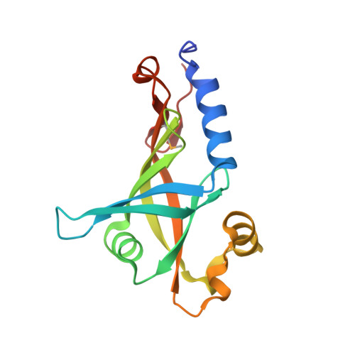 | |
UniProt | |||||
Find proteins for Q825J7 (Streptomyces avermitilis (strain ATCC 31267 / DSM 46492 / JCM 5070 / NBRC 14893 / NCIMB 12804 / NRRL 8165 / MA-4680)) Explore Q825J7 Go to UniProtKB: Q825J7 | |||||
Entity Groups | |||||
| Sequence Clusters | 30% Identity50% Identity70% Identity90% Identity95% Identity100% Identity | ||||
| UniProt Group | Q825J7 | ||||
Sequence AnnotationsExpand | |||||
| |||||
| Ligands 1 Unique | |||||
|---|---|---|---|---|---|
| ID | Chains | Name / Formula / InChI Key | 2D Diagram | 3D Interactions | |
| IPA Query on IPA | C [auth A], D [auth B], E [auth B], F [auth B] | ISOPROPYL ALCOHOL C3 H8 O KFZMGEQAYNKOFK-UHFFFAOYSA-N |  | ||
| Modified Residues 1 Unique | |||||
|---|---|---|---|---|---|
| ID | Chains | Type | Formula | 2D Diagram | Parent |
| MSE Query on MSE | A, B | L-PEPTIDE LINKING | C5 H11 N O2 Se |  | MET |
| Length ( Å ) | Angle ( ˚ ) |
|---|---|
| a = 87.89 | α = 90 |
| b = 87.89 | β = 90 |
| c = 151.58 | γ = 120 |
| Software Name | Purpose |
|---|---|
| MolProbity | model building |
| SOLVE | phasing |
| REFMAC | refinement |
| SCALEPACK | data scaling |
| PDB_EXTRACT | data extraction |
| DENZO | data reduction |
RCSB PDB (citation) is hosted by
RCSB PDB is a member of the

