


Crystal Structure of Plasmodium vivax 2-Cys Peroxiredoxin, Reduced
Artz, J.D., Qiu, W., Dong, A., Lew, J., Ren, H., Zhao, Y., Kozieradski, I., Edwards, A.M., Arrowsmith, C.H., Weigelt, J., Sundstrom, M., Bochkarev, A., Hui, R.To be published.
Experimental Data Snapshot
wwPDB Validation 3D Report Full Report

Entity ID: 1 | |||||
|---|---|---|---|---|---|
| Molecule | Chains | Sequence Length | Organism | Details | Image |
| 2-Cys Peroxiredoxin | 213 | Plasmodium vivax Sal-1 | Mutation(s): 0 | 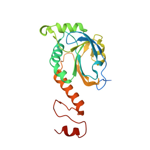 | |
UniProt | |||||
Find proteins for A5K421 (Plasmodium vivax (strain Salvador I)) Explore A5K421 Go to UniProtKB: A5K421 | |||||
Entity Groups | |||||
| Sequence Clusters | 30% Identity50% Identity70% Identity90% Identity95% Identity100% Identity | ||||
| UniProt Group | A5K421 | ||||
Sequence AnnotationsExpand | |||||
| |||||
| Length ( Å ) | Angle ( ˚ ) |
|---|---|
| a = 91.352 | α = 90 |
| b = 212.575 | β = 90 |
| c = 115.261 | γ = 90 |
| Software Name | Purpose |
|---|---|
| HKL-2000 | data collection |
| MOLREP | phasing |
| REFMAC | refinement |
| HKL-2000 | data reduction |
| HKL-2000 | data scaling |
RCSB PDB (citation) is hosted by
RCSB PDB is a member of the

