2I7O

Structure of Re(4,7-dimethyl-phen)(Thr124His)(Lys122Trp)(His83Gln)AzCu(II), a Rhenium modified Azurin mutant


Experimental Data Snapshot

  • Method: X-RAY DIFFRACTION
  • Resolution: 1.50 Å
  • R-Value Free: 0.255 
  • R-Value Work: 0.236 

wwPDB Validation   3D Report Full Report


Ligand Structure Quality Assessment 


This is version 1.2 of the entry. See complete history


Literature

Tryptophan-accelerated electron flow through proteins.

Shih, C.Museth, A.K.Abrahamsson, M.Blanco-Rodriguez, A.M.Di Bilio, A.J.Sudhamsu, J.Crane, B.R.Ronayne, K.L.Towrie, M.Vlcek, A.Richards, J.H.Winkler, J.R.Gray, H.B.

(2008) Science 320: 1760-1762

  • DOI: https://doi.org/10.1126/science.1158241
  • Primary Citation of Related Structures:  
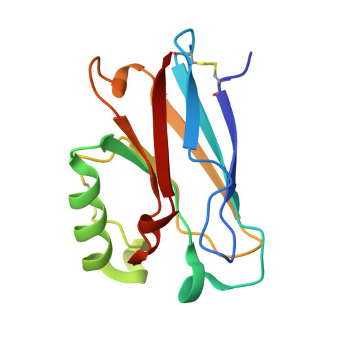
    2I7O

  • PubMed Abstract: 

    Energy flow in biological structures often requires submillisecond charge transport over long molecular distances. Kinetics modeling suggests that charge-transfer rates can be greatly enhanced by multistep electron tunneling in which redox-active amino acid side chains act as intermediate donors or acceptors. We report transient optical and infrared spectroscopic experiments that quantify the extent to which an intervening tryptophan residue can facilitate electron transfer between distant metal redox centers in a mutant Pseudomonas aeruginosa azurin. Cu(I) oxidation by a photoexcited Re(I)-diimine at position 124 on a histidine(124)-glycine(123)-tryptophan(122)-methionine(121) beta strand occurs in a few nanoseconds, fully two orders of magnitude faster than documented for single-step electron tunneling at a 19 angstrom donor-acceptor distance.


  • Organizational Affiliation

    Beckman Institute, California Institute of Technology, Pasadena, CA 91125, USA.


Macromolecules
Find similar proteins by:  (by identity cutoff)  |  3D Structure
Entity ID: 1
MoleculeChains Sequence LengthOrganismDetailsImage
Azurin128Pseudomonas aeruginosaMutation(s): 3 
Gene Names: azu
UniProt
Find proteins for P00282 (Pseudomonas aeruginosa (strain ATCC 15692 / DSM 22644 / CIP 104116 / JCM 14847 / LMG 12228 / 1C / PRS 101 / PAO1))
Explore P00282 
Go to UniProtKB:  P00282
Entity Groups  
Sequence Clusters30% Identity50% Identity70% Identity90% Identity95% Identity100% Identity
UniProt GroupP00282
Sequence Annotations
Expand
  • Reference Sequence
Small Molecules
Ligands 2 Unique
IDChains Name / Formula / InChI Key2D Diagram3D Interactions
REQ
Query on REQ

Download Ideal Coordinates CCD File 
C [auth A](1,10 PHENANTHROLINE)-(TRI-CARBON MONOXIDE) RHENIUM (I)
C17 H12 N2 O3 Re
SINYNGNXCUIFTB-UHFFFAOYSA-N
CU
Query on CU

Download Ideal Coordinates CCD File 
B [auth A]COPPER (II) ION
Cu
JPVYNHNXODAKFH-UHFFFAOYSA-N
Experimental Data & Validation

Experimental Data

  • Method: X-RAY DIFFRACTION
  • Resolution: 1.50 Å
  • R-Value Free: 0.255 
  • R-Value Work: 0.236 
  • Space Group: I 2 2 2
Unit Cell:
Length ( Å )Angle ( ˚ )
a = 63.223α = 90
b = 69.075β = 90
c = 68.944γ = 90
Software Package:
Software NamePurpose
CNSrefinement
PDB_EXTRACTdata extraction
ADSCdata collection
HKL-2000data reduction
HKL-2000data scaling
AMoREphasing

Structure Validation

View Full Validation Report


Ligand Structure Quality Assessment 


Entry History 

Deposition Data

Revision History  (Full details and data files)

  • Version 1.0: 2007-08-14
    Type: Initial release
  • Version 1.1: 2011-07-13
    Changes: Version format compliance
  • Version 1.2: 2021-10-20
    Changes: Database references, Derived calculations