2I7F

Sphingomonas yanoikuyae B1 ferredoxin


Experimental Data Snapshot

  • Method: X-RAY DIFFRACTION
  • Resolution: 1.90 Å
  • R-Value Free: 0.240 
  • R-Value Work: 0.193 
  • R-Value Observed: 0.196 

wwPDB Validation   3D Report Full Report


This is version 1.4 of the entry. See complete history


Literature

Structural investigations of the ferredoxin and terminal oxygenase components of the biphenyl 2,3-dioxygenase from Sphingobium yanoikuyae B1.

Ferraro, D.J.Brown, E.N.Yu, C.L.Parales, R.E.Gibson, D.T.Ramaswamy, S.

(2007) BMC Struct Biol 7: 10-10

  • DOI: https://doi.org/10.1186/1472-6807-7-10
  • Primary Citation of Related Structures:  
    2GBW, 2GBX, 2I7F

  • PubMed Abstract: 

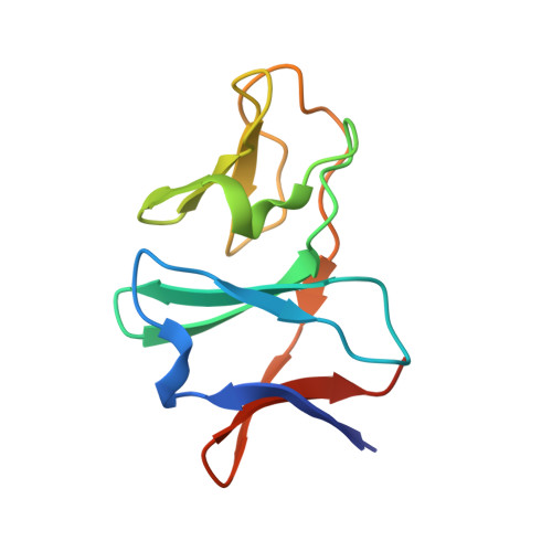
    The initial step involved in oxidative hydroxylation of monoaromatic and polyaromatic compounds by the microorganism Sphingobium yanoikuyae strain B1 (B1), previously known as Sphingomonas yanoikuyae strain B1 and Beijerinckia sp. strain B1, is performed by a set of multiple terminal Rieske non-heme iron oxygenases. These enzymes share a single electron donor system consisting of a reductase and a ferredoxin (BPDO-FB1). One of the terminal Rieske oxygenases, biphenyl 2,3-dioxygenase (BPDO-OB1), is responsible for B1's ability to dihydroxylate large aromatic compounds, such as chrysene and benzo[a]pyrene.


  • Organizational Affiliation

    Department of Biochemistry, University of Iowa Roy J. and Lucille A. Carver College of Medicine, Iowa City, Iowa, 52242, USA. daniel-ferraro@uiowa.edu


Macromolecules
Find similar proteins by:  (by identity cutoff)  |  3D Structure
Entity ID: 1
MoleculeChains Sequence LengthOrganismDetailsImage
Ferredoxin component of dioxygenase
A, B
108Sphingobium yanoikuyaeMutation(s): 0 
Gene Names: ahdA3
UniProt
Find proteins for A2TC31 (Sphingobium yanoikuyae)
Explore A2TC31 
Go to UniProtKB:  A2TC31
Entity Groups  
Sequence Clusters30% Identity50% Identity70% Identity90% Identity95% Identity100% Identity
UniProt GroupA2TC31
Sequence Annotations
Expand
  • Reference Sequence
Experimental Data & Validation

Experimental Data

  • Method: X-RAY DIFFRACTION
  • Resolution: 1.90 Å
  • R-Value Free: 0.240 
  • R-Value Work: 0.193 
  • R-Value Observed: 0.196 
  • Space Group: P 65 2 2
Unit Cell:
Length ( Å )Angle ( ˚ )
a = 62.064α = 90
b = 62.064β = 90
c = 238.436γ = 120
Software Package:
Software NamePurpose
d*TREKdata scaling
AMoREphasing
REFMACrefinement
PDB_EXTRACTdata extraction
MAR345data collection
d*TREKdata reduction

Structure Validation

View Full Validation Report


Entry History 

Deposition Data

Revision History  (Full details and data files)

  • Version 1.0: 2007-03-20
    Type: Initial release
  • Version 1.1: 2008-05-01
    Changes: Version format compliance
  • Version 1.2: 2011-07-13
    Changes: Version format compliance
  • Version 1.3: 2017-10-18
    Changes: Refinement description
  • Version 1.4: 2023-08-30
    Changes: Data collection, Database references, Derived calculations, Refinement description