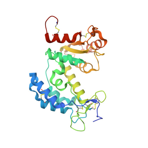
2I67

Structural Basis for the Mechanistic Understanding Human CD38 Controlled Multiple Catalysis


Experimental Data Snapshot

  • Method: X-RAY DIFFRACTION
  • Resolution: 1.71 Å
  • R-Value Free: 0.236 
  • R-Value Work: 0.189 
  • R-Value Observed: 0.191 

wwPDB Validation   3D Report Full Report


Ligand Structure Quality Assessment 


This is version 1.4 of the entry. See complete history


Literature

Structural basis for the mechanistic understanding of human CD38-controlled multiple catalysis.

Liu, Q.Kriksunov, I.A.Graeff, R.Munshi, C.Lee, H.C.Hao, Q.

(2006) J Biol Chem 281: 32861-32869

  • DOI: https://doi.org/10.1074/jbc.M606365200
  • Primary Citation of Related Structures:  
    2I65, 2I66, 2I67

  • PubMed Abstract: 

    The enzymatic cleavage of the nicotinamide-glycosidic bond on nicotinamide adenine dinucleotide (NAD(+)) has been proposed to go through an oxocarbenium ion-like transition state. Because of the instability of the ionic intermediate, there has been no structural report on such a transient reactive species. Human CD38 is an ectoenzyme that can use NAD(+) to synthesize two calcium-mobilizing molecules. By using NAD(+) and a surrogate substrate, NGD(+), we captured and determined crystal structures of the enzyme complexed with an intermediate, a substrate, and a product along the reaction pathway. Our results showed that the intermediate is stabilized by polar interactions with the catalytic residue Glu(226) rather than by a covalent linkage. The polar interactions between Glu(226) and the substrate 2',3'-OH groups are essential for initiating catalysis. Ser(193) was demonstrated to have a regulative role during catalysis and is likely to be involved in intermediate stabilization. In addition, a product inhibition effect by ADP-ribose (through the reorientation of the product) or GDP-ribose (through the formation of a covalently linked GDP-ribose dimer) was observed. These structural data provide insights into the understanding of multiple catalysis and clues for drug design.


  • Organizational Affiliation

    Macromolecular Diffraction Facility at the Cornell High Energy Synchrotron Source (MacCHESS), Cornell University, Ithaca, NY 14853, USA.


Macromolecules
Find similar proteins by:  (by identity cutoff)  |  3D Structure
Entity ID: 1
MoleculeChains Sequence LengthOrganismDetailsImage
ADP-ribosyl cyclase 1
A, B
262Homo sapiensMutation(s): 5 
Gene Names: CD38
EC: 3.2.2.5
UniProt & NIH Common Fund Data Resources
Find proteins for P28907 (Homo sapiens)
Explore P28907 
Go to UniProtKB:  P28907
PHAROS:  P28907
GTEx:  ENSG00000004468 
Entity Groups  
Sequence Clusters30% Identity50% Identity70% Identity90% Identity95% Identity100% Identity
UniProt GroupP28907
Sequence Annotations
Expand
  • Reference Sequence
Experimental Data & Validation

Experimental Data

  • Method: X-RAY DIFFRACTION
  • Resolution: 1.71 Å
  • R-Value Free: 0.236 
  • R-Value Work: 0.189 
  • R-Value Observed: 0.191 
  • Space Group: P 1
Unit Cell:
Length ( Å )Angle ( ˚ )
a = 41.893α = 109.36
b = 51.152β = 91.22
c = 65.639γ = 97.38
Software Package:
Software NamePurpose
REFMACrefinement
ADSCdata collection
HKL-2000data reduction
HKL-2000data scaling
MOLREPphasing

Structure Validation

View Full Validation Report


Ligand Structure Quality Assessment 


Entry History 

Deposition Data

Revision History  (Full details and data files)

  • Version 1.0: 2006-09-05
    Type: Initial release
  • Version 1.1: 2008-05-01
    Changes: Version format compliance
  • Version 1.2: 2011-07-13
    Changes: Version format compliance
  • Version 1.3: 2021-10-20
    Changes: Database references, Derived calculations
  • Version 1.4: 2023-08-30
    Changes: Data collection, Refinement description