2I5X

Engineering the PTPbeta catalytic domain with improved crystallization properties


Experimental Data Snapshot

  • Method: X-RAY DIFFRACTION
  • Resolution: 1.70 Å
  • R-Value Free: 0.207 
  • R-Value Work: 0.179 
  • R-Value Observed: 0.180 

wwPDB Validation   3D Report Full Report


Ligand Structure Quality Assessment 


This is version 1.3 of the entry. See complete history


Literature

Engineering the catalytic domain of human protein tyrosine phosphatase beta for structure-based drug discovery.

Evdokimov, A.G.Pokross, M.Walter, R.Mekel, M.Cox, B.Li, C.Bechard, R.Genbauffe, F.Andrews, R.Diven, C.Howard, B.Rastogi, V.Gray, J.Maier, M.Peters, K.G.

(2006) Acta Crystallogr D Biol Crystallogr 62: 1435-1445

  • DOI: https://doi.org/10.1107/S0907444906037784
  • Primary Citation of Related Structures:  
    2HC1, 2HC2, 2I3R, 2I3U, 2I4E, 2I4G, 2I4H, 2I5X

  • PubMed Abstract: 

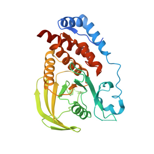
    Protein tyrosine phosphatases (PTPs) play roles in many biological processes and are considered to be important targets for drug discovery. As inhibitor development has proven challenging, crystal structure-based design will be very helpful to advance inhibitor potency and selectivity. Successful application of protein crystallography to drug discovery heavily relies on high-quality crystal structures of the protein of interest complexed with pharmaceutically interesting ligands. It is very important to be able to produce protein-ligand crystals rapidly and reproducibly for as many ligands as necessary. This study details our efforts to engineer the catalytic domain of human protein tyrosine phosphatase beta (HPTPbeta-CD) with properties suitable for rapid-turnaround crystallography. Structures of apo HPTPbeta-CD and its complexes with several novel small-molecule inhibitors are presented here for the first time.


  • Organizational Affiliation

    Structural Biology, Procter and Gamble Pharmaceuticals, 8700 Mason-Montgomery Road, Mason, OH 45140, USA. artem@xtals.org


Macromolecules
Find similar proteins by:  (by identity cutoff)  |  3D Structure
Entity ID: 1
MoleculeChains Sequence LengthOrganismDetailsImage
Receptor-type tyrosine-protein phosphatase beta
A, B
313Homo sapiensMutation(s): 0 
Gene Names: PTPRBPTPB
EC: 3.1.3.48
UniProt & NIH Common Fund Data Resources
Find proteins for P23467 (Homo sapiens)
Explore P23467 
Go to UniProtKB:  P23467
PHAROS:  P23467
GTEx:  ENSG00000127329 
Entity Groups  
Sequence Clusters30% Identity50% Identity70% Identity90% Identity95% Identity100% Identity
UniProt GroupP23467
Sequence Annotations
Expand
  • Reference Sequence
Experimental Data & Validation

Experimental Data

  • Method: X-RAY DIFFRACTION
  • Resolution: 1.70 Å
  • R-Value Free: 0.207 
  • R-Value Work: 0.179 
  • R-Value Observed: 0.180 
  • Space Group: P 1 21 1
Unit Cell:
Length ( Å )Angle ( ˚ )
a = 61.973α = 90
b = 71.308β = 93.4
c = 69.69γ = 90
Software Package:
Software NamePurpose
REFMACrefinement
PDB_EXTRACTdata extraction
HKL-2000data collection
HKL-2000data reduction
HKL-2000data scaling
AMoREphasing

Structure Validation

View Full Validation Report


Ligand Structure Quality Assessment 


Entry History 

Deposition Data

Revision History  (Full details and data files)

  • Version 1.0: 2006-09-05
    Type: Initial release
  • Version 1.1: 2008-05-01
    Changes: Version format compliance
  • Version 1.2: 2011-07-13
    Changes: Advisory, Version format compliance
  • Version 1.3: 2023-08-30
    Changes: Data collection, Database references, Derived calculations, Refinement description