2I5P

Crystal structure of glyceraldehyde-3-phosphate dehydrogenase isoform 1 from K. marxianus


Experimental Data Snapshot

  • Method: X-RAY DIFFRACTION
  • Resolution: 2.30 Å
  • R-Value Free: 0.245 
  • R-Value Work: 0.216 
  • R-Value Observed: 0.216 

wwPDB Validation   3D Report Full Report


Ligand Structure Quality Assessment 


This is version 1.5 of the entry. See complete history


Literature

The Crystal and Solution Structures of Glyceraldehyde-3-phosphate Dehydrogenase Reveal Different Quaternary Structures.

Ferreira-da-Silva, F.Pereira, P.J.Gales, L.Roessle, M.Svergun, D.I.Moradas-Ferreira, P.Damas, A.M.

(2006) J Biol Chem 281: 33433-33440

  • DOI: https://doi.org/10.1074/jbc.M605267200
  • Primary Citation of Related Structures:  
    2I5P

  • PubMed Abstract: 

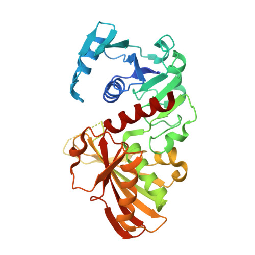
    The presence of an isoform of glyceraldehyde-3-phosphate dehydrogenase (kmGAPDH1p) associated with the cell wall of a flocculent strain of Kluyveromyces marxianus was the first report of a non-cytosolic localization of a glycolytic enzyme, but the mechanism by which the protein is transported to the cell surface is not known. To identify structural features that could account for the multiple localizations of the protein, the three-dimensional structure of kmGAPDH1p was determined by x-ray crystallography and small angle x-ray scattering. The x-ray crystallographic structure of kmGAPDH1p revealed a dimer, although all GAPDH homologs studied thus far have a tetrameric structure with 222 symmetry. Interestingly, the structure of kmGAPDH1p in solution revealed a tetramer with a 70 degrees tilt angle between the dimers. Moreover, the separation between the centers of the dimers composing the kmGAPDH1p tetramer diminished from 34 to 30 A upon NAD(+) binding, this latter value being similar to the observed in the crystallographic models of GAPDH homologs. The less compact structure of apo-kmGAPDH1p could already be the first image of the transition intermediate between the tetramer observed in solution and the dimeric form found in the crystal structure, which we postulate to exist in vivo because of the protein's multiple subcellular localizations in this yeast species.


  • Organizational Affiliation

    Instituto de Biologia Molecular e Celular, Universidade do Porto, 4150-180 Porto, Portugal.


Macromolecules
Find similar proteins by:  (by identity cutoff)  |  3D Structure
Entity ID: 1
MoleculeChains Sequence LengthOrganismDetailsImage
Glyceraldehyde-3-phosphate dehydrogenase 1A [auth O],
B [auth P]
342Kluyveromyces marxianusMutation(s): 0 
Gene Names: GAP1
EC: 1.2.1.12
UniProt
Find proteins for P84998 (Kluyveromyces marxianus)
Explore P84998 
Go to UniProtKB:  P84998
Entity Groups  
Sequence Clusters30% Identity50% Identity70% Identity90% Identity95% Identity100% Identity
UniProt GroupP84998
Sequence Annotations
Expand
  • Reference Sequence
Experimental Data & Validation

Experimental Data

  • Method: X-RAY DIFFRACTION
  • Resolution: 2.30 Å
  • R-Value Free: 0.245 
  • R-Value Work: 0.216 
  • R-Value Observed: 0.216 
  • Space Group: P 41 21 2
Unit Cell:
Length ( Å )Angle ( ˚ )
a = 70.9α = 90
b = 70.9β = 90
c = 358.4γ = 90
Software Package:
Software NamePurpose
ADSCdata collection
AMoREphasing
CNSrefinement
MOSFLMdata reduction
CCP4data scaling

Structure Validation

View Full Validation Report


Ligand Structure Quality Assessment 


Entry History 

Deposition Data

Revision History  (Full details and data files)

  • Version 1.0: 2006-09-12
    Type: Initial release
  • Version 1.1: 2008-05-01
    Changes: Version format compliance
  • Version 1.2: 2011-07-13
    Changes: Version format compliance
  • Version 1.3: 2020-04-15
    Changes: Advisory, Data collection, Database references
  • Version 1.4: 2020-07-29
    Type: Remediation
    Reason: Carbohydrate remediation
    Changes: Data collection, Derived calculations, Structure summary
  • Version 1.5: 2023-08-30
    Changes: Advisory, Data collection, Database references, Refinement description, Structure summary