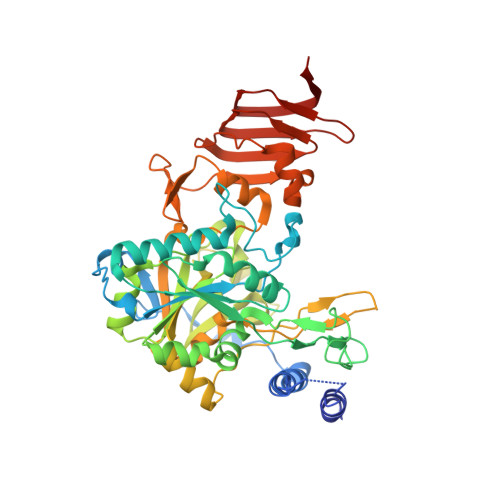
Structural Basis for Subunit Assembly in UDP-glucose Pyrophosphorylase from Saccharomyces cerevisiae
Roeben, A., Plitzko, J.M., Koerner, R., Boettcher, U.M.K., Siegers, K., Hayer-Hartl, M., Bracher, A.(2006) J Mol Biol 364: 551-560
- PubMed: 17010990
- DOI: https://doi.org/10.1016/j.jmb.2006.08.079
- Primary Citation of Related Structures:
2I5K - PubMed Abstract:
UDP-glucose is the universal activated form of glucose, employed in all organisms for glucosyl transfer reactions and as precursor for various activated carbohydrates. In animal and fungal metabolism, UDP-glucose is required for utilization of galactose and for the synthesis of glycogen, the major carbohydrate storage polymer. The formation of UDP-glucose is catalyzed by UDP-glucose pyrophosphorylase (UGPase), which is highly conserved among eukaryotes. Here, we present the crystal structure of yeast UGPase, Ugp1p. Both in solution and in the crystal, Ugp1p forms homooctamers, which represent the enzymatically active form of the protein. Ugp1p subunits consist of three domains, with the active site presumably located in the central SpsA GnT I core (SGC) domain. The association in the octamer is mediated by contacts between left-handed beta-helices in the C-terminal domains, forming a toroidal solenoid structure in the core of the complex. The catalytic domains attached to this scaffold core do not directly contact each other, consistent with simple Michaelis-Menten kinetics found for Ugp1p. Conservation of hydrophobic residues at the subunit interfaces suggests that all fungal and animal homologs form this quarternary structure arrangement in contrast to monomeric plant UGPases, which have charged residues at these positions. Implications of this oligomeric arrangement for regulation of UGPase activity in fungi and animals are discussed.
Organizational Affiliation:
Department of Cellular Biochemistry, Max-Planck-Institute of Biochemistry, Am Klopferspitz 18, 82152 Martinsried, Germany.