2I3V

Measurement of conformational changes accompanying desensitization in an ionotropic glutamate receptor: Structure of G725C mutant


Experimental Data Snapshot

  • Method: X-RAY DIFFRACTION
  • Resolution: 2.40 Å
  • R-Value Free: 0.281 
  • R-Value Work: 0.237 
  • R-Value Observed: 0.237 

wwPDB Validation   3D Report Full Report


This is version 1.4 of the entry. See complete history


Literature

Measurement of Conformational Changes accompanying Desensitization in an Ionotropic Glutamate Receptor.

Armstrong, N.Jasti, J.Beich-Frandsen, M.Gouaux, E.

(2006) Cell 127: 85-97

  • DOI: https://doi.org/10.1016/j.cell.2006.08.037
  • Primary Citation of Related Structures:  
    2I3V, 2I3W

  • PubMed Abstract: 

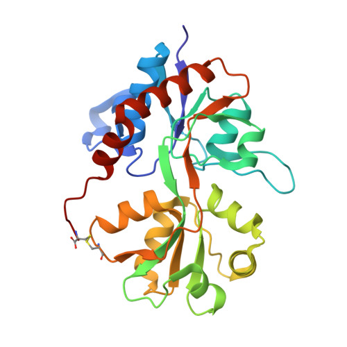
    The canonical conformational states occupied by most ligand-gated ion channels, and many cell-surface receptors, are the resting, activated, and desensitized states. While the resting and activated states of multiple receptors are well characterized, elaboration of the structural properties of the desensitized state, a state that is by definition inactive, has proven difficult. Here we use electrical, chemical, and crystallographic experiments on the AMPA-sensitive GluR2 receptor, defining the conformational rearrangements of the agonist binding cores that occur upon desensitization of this ligand-gated ion channel. These studies demonstrate that desensitization involves the rupture of an extensive interface between domain 1 of 2-fold related glutamate-binding core subunits, compensating for the ca. 21 degrees of domain closure induced by glutamate binding. The rupture of the domain 1 interface allows the ion channel to close and thereby provides a simple explanation to the long-standing question of how agonist binding is decoupled from ion channel gating upon receptor desensitization.


  • Organizational Affiliation

    Department of Biochemistry and Molecular Biophysics, Columbia University, 650 West 168th Street, New York, NY 10032 USA.


Macromolecules
Find similar proteins by:  (by identity cutoff)  |  3D Structure
Entity ID: 1
MoleculeChains Sequence LengthOrganismDetailsImage
Glutamate receptor 2
A, B, C, D
259Rattus norvegicusMutation(s): 0 
Gene Names: Gria2Glur2
UniProt
Find proteins for P19491 (Rattus norvegicus)
Explore P19491 
Go to UniProtKB:  P19491
Entity Groups  
Sequence Clusters30% Identity50% Identity70% Identity90% Identity95% Identity100% Identity
UniProt GroupP19491
Sequence Annotations
Expand
  • Reference Sequence
Small Molecules
Ligands 2 Unique
IDChains Name / Formula / InChI Key2D Diagram3D Interactions
GLU
Query on GLU

Download Ideal Coordinates CCD File 
G [auth A],
K [auth B],
M [auth C],
O [auth D]
GLUTAMIC ACID
C5 H9 N O4
WHUUTDBJXJRKMK-VKHMYHEASA-N
ZN
Query on ZN

Download Ideal Coordinates CCD File 
E [auth A]
F [auth A]
H [auth B]
I [auth B]
J [auth B]
E [auth A],
F [auth A],
H [auth B],
I [auth B],
J [auth B],
L [auth C],
N [auth D]
ZINC ION
Zn
PTFCDOFLOPIGGS-UHFFFAOYSA-N
Binding Affinity Annotations 
IDSourceBinding Affinity
GLU PDBBind:  2I3V Ki: 282 (nM) from 1 assay(s)
Experimental Data & Validation

Experimental Data

  • Method: X-RAY DIFFRACTION
  • Resolution: 2.40 Å
  • R-Value Free: 0.281 
  • R-Value Work: 0.237 
  • R-Value Observed: 0.237 
  • Space Group: P 21 21 21
Unit Cell:
Length ( Å )Angle ( ˚ )
a = 63.191α = 90
b = 106.291β = 90
c = 192.817γ = 90
Software Package:
Software NamePurpose
CNSrefinement
PDB_EXTRACTdata extraction
HKL-2000data reduction
HKL-2000data scaling
AMoREphasing

Structure Validation

View Full Validation Report


Entry History 

Deposition Data

Revision History  (Full details and data files)

  • Version 1.0: 2006-10-17
    Type: Initial release
  • Version 1.1: 2008-05-01
    Changes: Version format compliance
  • Version 1.2: 2011-07-13
    Changes: Version format compliance
  • Version 1.3: 2017-08-16
    Changes: Refinement description, Source and taxonomy
  • Version 1.4: 2023-08-30
    Changes: Data collection, Database references, Derived calculations, Refinement description